Microbes in Manhattan

October 1918: The Story of Nurse Battey

The Great War and the Terrible Flu

In the last months of World War I a new influenza swept the world. In Manhattan it filled the college with sick soldiers, and a young Red Cross nurse gave her life caring for them.

How Manhattan bought a machine to breathe for its children

Iron Lung, Iron Will

In 1937 the American Legion asked Manhattan to buy an iron lung. Clubs, schoolchildren, a dance and even a wolf hunt raised $1,249.50, and the machine stood ready at the fire station for more than a decade.

Fear, resilience and a vaccine

The Polio Epidemic

For half a century, polio returned every summer. It reshaped families, hospitals, philanthropy and the way we design the world around us.

How vaccines conquered smallpox and nearly ended polio

Immunization

From the ancient practice of variolation to Jenner’s cowpox and the polio vaccines of the 1950s, vaccines have saved more lives than almost any other invention. Explore how they work, what they prevent, and the long road to a polio-free world.

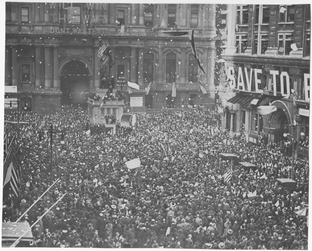
A vast crowd fills a city street under flags and confetti at the first news of peace, November 11, 1918.
Influenza has been able to kill more American soldier boys than all the kaiser’s troops and submarines.

The Riley County Chronicle, October 15, 1918

Introduction

The 1918 influenza pandemic was the most severe pandemic in recent history. Arriving during the final months of World War I, the H1N1 virus of avian origin killed more people in twelve weeks than the war had in four years.

Although there is not universal consensus regarding where the virus originated, it created a worldwide crisis from 1918 to 1919.

A vast crowd fills a city street under flags and confetti at the first news of peace, November 11, 1918.
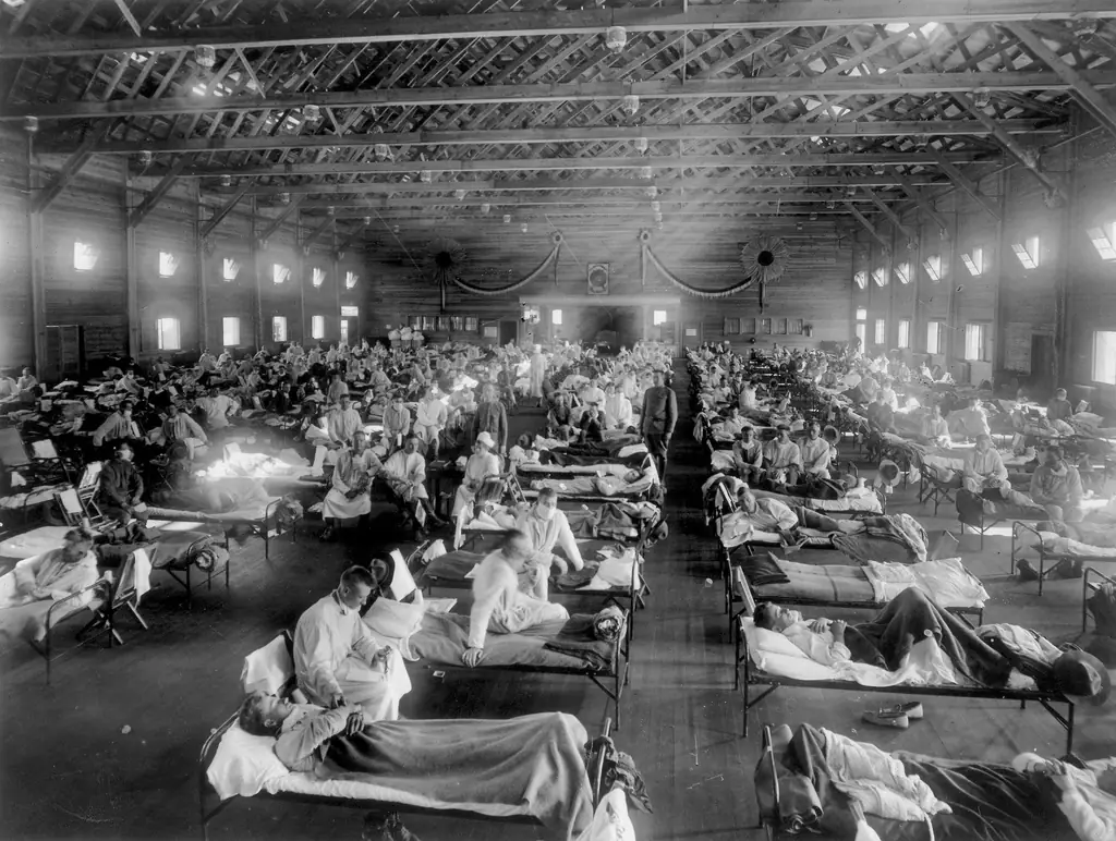
Long rows of beds in an emergency hospital ward at Camp Funston, Fort Riley, Kansas, 1918.
Spring 1918

Influenza was a known disease in 1918, but it was traditionally a mild illness with symptoms departing after a few days. Seasonal outbreaks occurred annually with generally minor effects. Just as in prior years, in the spring of 1918 the flu swept through many U.S. Army training camps.

As a result of this seasonal outbreak, Camp Funston at Fort Riley, near Manhattan, Kansas, eventually became the target of rumors. These rumors led some researchers to place the spring outbreak as the beginning of the much more catastrophic one that came in the fall.

However, despite a higher than average number of cases being reported, few of the patients infected in the spring succumbed to the illness. This has led some researchers, such as Jed Dunham, to declare the spring event an entirely independent bout of seasonal flu, unrelated to the fall pandemic which took millions of lives. Clearing this misconception would also clear Camp Funston as the starting point of the infection.

Long rows of beds in an emergency hospital ward at Camp Funston, Fort Riley, Kansas, 1918.
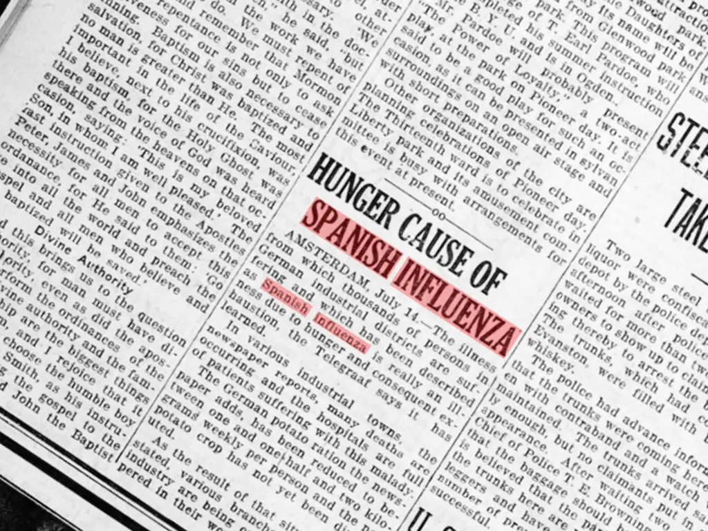
A 1918 newspaper column with the headline “Hunger Cause of Spanish Influenza.”

Naming the Illness

The seasonal flu which began in North America in the spring may have been taken to Europe by American soldiers on their way to the war. Once it arrived in France, it spread rapidly.

Largely unrestricted and unaffected by the ongoing war, Spain was the primary country reporting on the spread of the highly contagious seasonal flu in Europe. As in North America that spring, there was a higher than average number of flu cases reported, and a low number of deaths.

As newspapers in the U.S. began to write stories about this illness spreading in Spain and the rest of Europe, they began calling it the “Spanish Flu.” The name became ingrained in the public mind in the following months, as the separate pandemic strain spread in the fall.

A 1918 newspaper column with the headline “Hunger Cause of Spanish Influenza.”

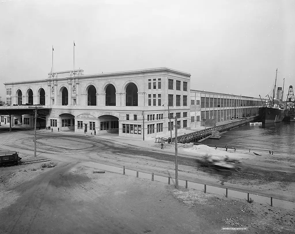
Commonwealth Pier near Boston, Massachusetts, a long arched pier building on the waterfront.

Summer 1918

Around August 27, sailors stationed on board the Receiving Ship at Commonwealth Pier in Boston began reporting to sick bay with the usual symptoms of influenza. By August 30, over 60 sailors were sick.

Soon Commonwealth Pier was overwhelmed, and 50 cases had to be transferred to Chelsea Naval Hospital. Flu sufferers commonly described feeling as if they had been beaten all over with a club.

This sickness had most of the common symptoms of the known influenza, but it seemed to bring much stronger reactions from patients, and many ultimately died. It was unlike any flu that had come before.

Commonwealth Pier near Boston, Massachusetts, a long arched pier building on the waterfront.

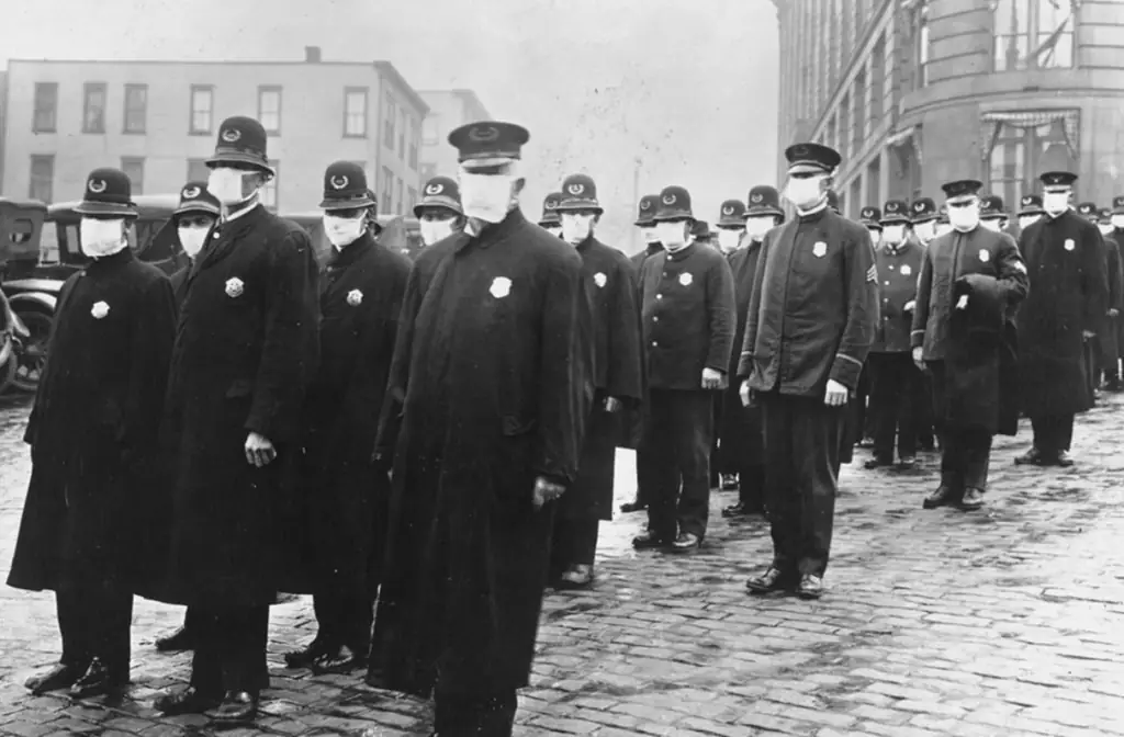
A line of police officers in long coats and caps wearing white gauze masks, 1918.
What Is a Pandemic?

Most people are sickened by influenza by the age of five. If a person survives (most flu deaths traditionally strike the very young or the very old), that person develops immunities which help to combat future arrivals of the seasonal illness.

A pandemic occurs when a new version of the disease evolves for which there are no known immunities. This is what happened in 1918.
Who died of influenza and pneumonia in the United States
01,0002,0003,000Deaths per 100,000<11–45–1415–2425–3435–4445–5455–6465–7475–8485+AgeAge <1: 1,500 per 100,000Age 1–4: 254 per 100,000Age 5–14: 18 per 100,000Age 15–24: 27 per 100,000Age 25–34: 45 per 100,000Age 35–44: 72 per 100,000Age 45–54: 127 per 100,000Age 55–64: 236 per 100,000Age 65–74: 555 per 100,000Age 75–84: 1,327 per 100,000Age 85+: 2,809 per 100,000Age <1: 2,255 per 100,000Age 1–4: 718 per 100,000Age 5–14: 173 per 100,000Age 15–24: 573 per 100,000Age 25–34: 991 per 100,000Age 35–44: 555 per 100,000Age 45–54: 345 per 100,000Age 55–64: 382 per 100,000Age 65–74: 645 per 100,000Age 75–84: 1,182 per 100,000Age 85+: 2,245 per 100,000Ages 25–34: 22× the usual rate
  • A usual year (1911–1917 average)
  • 1918

In a usual year, flu killed mostly the very young and the very old. In 1918 it also killed young adults in their prime. Source: CDC.

The 1918 pandemic struck people aged 25 to 40 the hardest. Modern medical science has yet to definitively answer why, but it seems plausible that the flu this age group was exposed to in childhood (1890 to 1900) was different from the H1N1 influenza which erupted in 1918.

Arriving with a cough or a sneeze, within days it could plunge a community into a nightmare of catastrophic despair.

A line of police officers in long coats and caps wearing white gauze masks, 1918.
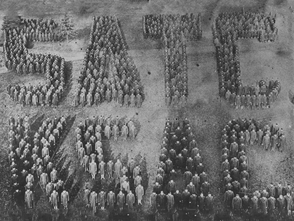
Kansas State Agricultural College Student Army Training Corps standing in formation to spell out letters, Manhattan, Kansas.
The Story of Nurse Battey

Surpassing any possibility of containment, by October the pandemic had reached Manhattan, Kansas. The available resources in the community were rapidly overwhelmed.

Kansas State Agricultural College, today Kansas State University, was heavily affected. One group living on campus, the Student Army Training Corps, was met with the highly contagious pandemic strain. To handle the high number of patients, help was sought from outside the community. A nurse named Sarah Battey answered that request.

At the time, influenza was not recognized as a deadly disease. As people began dying of the new strain in large numbers, fear ran rampant. The pandemic struck rapidly, and was especially violent in the close quarters of the army camps and the barracks that housed the Student Army Training Corps in Manhattan.

Kansas State Agricultural College Student Army Training Corps standing in formation to spell out letters, Manhattan, Kansas.

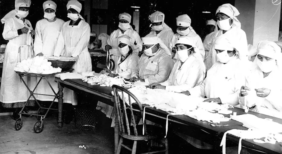
Red Cross volunteers in white gowns and gauze masks at long tables, Camp Devens, 1918.

Accounts by the American Red Cross

Red Cross nurses were sent wherever the epidemic broke out. This account comes from a Red Cross nurse caring for American soldiers at Base Hospital No. 57 in France.

The men were desperately sick. On the fourth night, the theater of this old college was opened to accommodate the great numbers. There were 63 new patients that night, besides the ones in my two tents and in the theater. I was told that at midnight more nurses would arrive to help me. Four of the doctors stayed on.

The men were the sickest I have ever seen. They looked like gassed patients but none of them had ever been near the front and they had only been in France three weeks, taking care of gassed horses. If ever doctors worked with patients, those from Base Hospital No. 57 certainly did, but those boys in spite of all our efforts simply died. One night 13 were carried out of my ward.

It was discouraging beyond all words to see those splendid specimens of manhood just pass out without a struggle. They were all big Western fellows; many of them had never had a sick day in their lives before.

A History of American Red Cross Nursing, 1922

Red Cross volunteers in white gowns and gauze masks at long tables, Camp Devens, 1918.

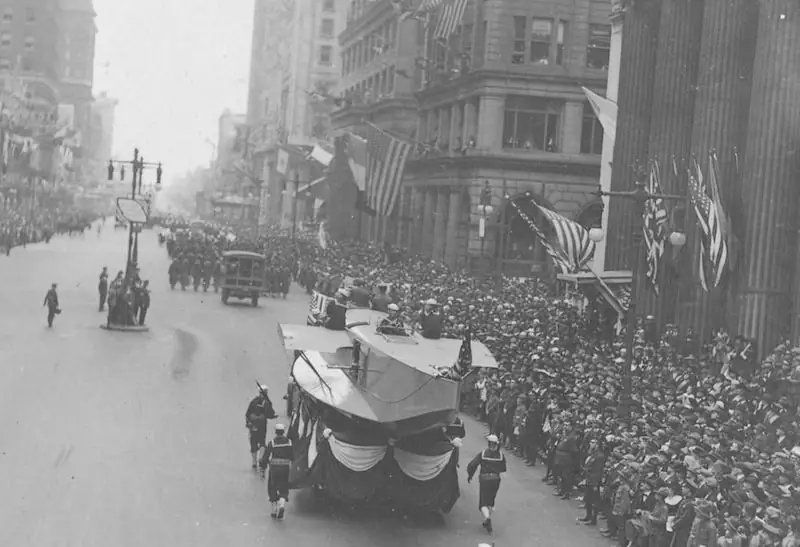
Crowds line a city street as a Liberty Loan parade passes, September 1918.

Proclamation to Close K.S.A.C.

On October 11, 1918, at the request of Governor Arthur Capper, the college announced it would close for a week. Students were asked to stay in Manhattan rather than travel home by train and spread the disease.

K. S. A. C. Proclamation

Complying with the proclamation and request of Governor Capper, College classes will be dismissed for the period of time beginning Saturday morning, October 12, ending with and including Saturday, October 19, this action having for its purpose the prevention of the spread of epidemic influenza within the State.

This statement does not apply to members of the Students’ Army Training Corps. They will receive orders from the Commandant, Captain Sturges.

The State Board of Health appeals to you, through me, to remain quietly in the city of Manhattan, not to go to your homes unless they are accessible by some other means than the railroad. To leave the city will endanger your own health and the health of others. The civilian population of Manhattan was never freer from disease than it is today. You are safer here during this period than anywhere else. Every measure will be taken to safeguard your health. The College physician will be at hand to render aid when called upon, which should be upon the first signs of indisposition.

Your action in the present emergency will be the test of your patriotism and public spirit.

W. M. Jardine, President. The Kansas State Collegian, October 11, 1918

Crowds line a city street as a Liberty Loan parade passes, September 1918.

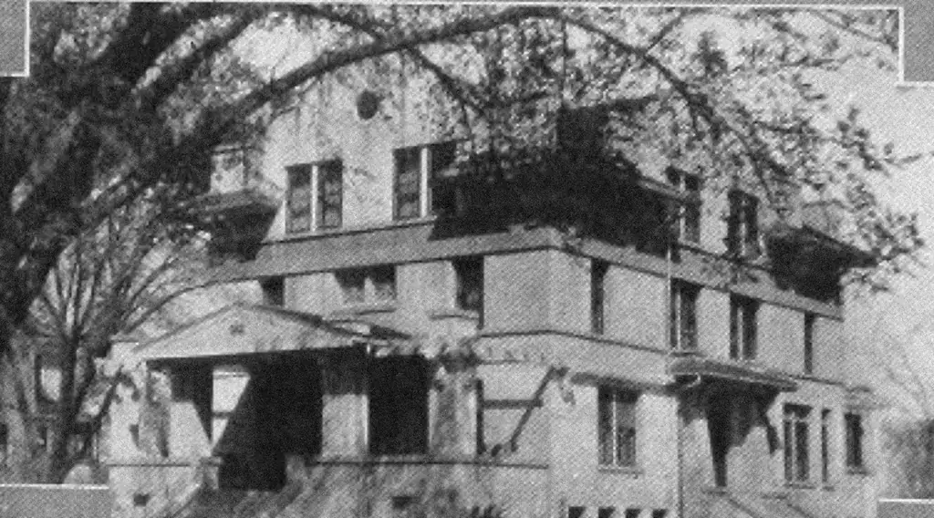
The old Sigma Alpha Epsilon fraternity house, a large three-story brick house behind bare trees.

SAE House Used as Temporary Hospital

Because of the overwhelming number of patients, many students were treated in buildings rapidly converted into hospitals. The YMCA and the Sigma Alpha Epsilon and Beta Theta Pi fraternity houses all took in the sick.

Sarah Battey volunteered to help, arriving on Monday, October 14, 1918. She reported to the Sigma Alpha Epsilon fraternity house, where many of the sick cadets were being treated.

By Saturday she had fallen ill herself. She died of the pandemic flu on October 19, 1918.

The old Sigma Alpha Epsilon fraternity house, a large three-story brick house behind bare trees.

Kansas State Agricultural College Student Army Training Corps in formation, Manhattan, Kansas.

A Formal Obituary

News of her death reached her husband’s family in Council Bluffs, Iowa, the same morning. She was buried there the next day.

Sarah H. Battey

H.V. Battey received word Saturday morning from Manhattan, Kansas announcing the death of Mrs. Sarah Battey, wife of Captain Percy B. Battey, of the medical corps at Camp Funston. Captain Battey is a son of Mr. and Mrs. H.V. Battey of this city. Mrs. Battey, who was formerly a trained nurse, had been living at Manhattan since her husband was attached to Camp Funston.

Monday evening she received a call to act as nurse for the students’ army training corps at the agricultural college at Manhattan, where influenza is prevalent. After several days of close attention to her work she was taken ill and passed away early Saturday morning.

The services were held at the grave in Fairview Cemetery. Rev. Wilford E. Mann, rector of St. Paul’s Episcopal Church, conducted the services.

Council Bluffs Daily Nonpareil, Sunday, October 20, 1918, page 8

She lies in Fairview Cemetery, Council Bluffs: Section J, Lot 65, Sarah Hillary Battey, March 19, 1890 – October 19, 1918.

Kansas State Agricultural College Student Army Training Corps in formation, Manhattan, Kansas.

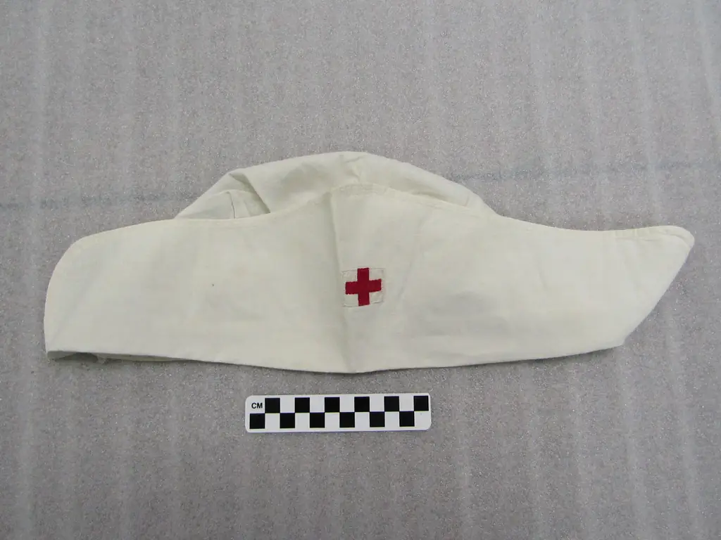
A white Red Cross nurse’s cap with a small red cross, from the Riley County Historical Museum collection.

Honoring the Fallen

Four days after her death, the college newspaper asked that she never be forgotten.

The College Owes Her Honor

The college should remember perpetually Mrs. Percy B. Battey, who gave her life for the sake of college students.

A temporary resident of Manhattan, with no ties to bind her to the state or to the college, Mrs. Battey, who was a trained nurse by profession, offered her services in caring for the young men who were most seriously ill with influenza. She contracted the disease, it passed into pneumonia, and after a few days she died.

Mrs. Battey made the supreme sacrifice in a cause in which no selfish interest could in any way have intruded. Hers was the pure spirit of sacrifice for humanity.

The Kansas Industrialist, October 23, 1918

A white Red Cross nurse’s cap with a small red cross, from the Riley County Historical Museum collection.

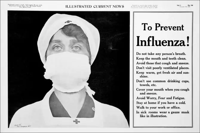
A 1918 poster headed “To Prevent Influenza!” beside a photograph of a nurse in a gauze mask.

From Tragedy Comes Knowledge

Millions of lives were lost in the 1918 influenza pandemic. As the average life expectancy dropped by 12 years, medical science was forced to modernize rapidly.

Weekly deaths from all causes, 1918 – 1919
0204060Deaths per 1,000 (annual rate)JuneJulyAug.Sep.Oct.Nov.Dec.Jan.Feb.Mar.1918 – 1919Oct. 19: Nurse Battey diesNew York, week of June 8: 12 per 1,000New York, week of June 15: 11 per 1,000New York, week of June 22: 11 per 1,000New York, week of June 29: 13 per 1,000New York, week of July 6: 11 per 1,000New York, week of July 13: 11 per 1,000New York, week of July 20: 11 per 1,000New York, week of July 27: 12 per 1,000New York, week of Aug. 3: 11 per 1,000New York, week of Aug. 10: 12 per 1,000New York, week of Aug. 17: 11 per 1,000New York, week of Aug. 24: 10 per 1,000New York, week of Aug. 31: 10 per 1,000New York, week of Sep. 7: 11 per 1,000New York, week of Sep. 14: 12 per 1,000New York, week of Sep. 21: 11 per 1,000New York, week of Sep. 28: 13 per 1,000New York, week of Oct. 5: 18 per 1,000New York, week of Oct. 12: 31 per 1,000New York, week of Oct. 19: 54 per 1,000New York, week of Oct. 26: 63 per 1,000New York, week of Nov. 2: 53 per 1,000New York, week of Nov. 9: 37 per 1,000New York, week of Nov. 16: 24 per 1,000New York, week of Nov. 23: 19 per 1,000New York, week of Nov. 30: 17 per 1,000New York, week of Dec. 7: 18 per 1,000New York, week of Dec. 14: 18 per 1,000New York, week of Dec. 21: 19 per 1,000New York, week of Dec. 28: 20 per 1,000New York, week of Jan. 4: 21 per 1,000New York, week of Jan. 11: 23 per 1,000New York, week of Jan. 18: 26 per 1,000New York, week of Jan. 25: 28 per 1,000London, week of June 8: 11 per 1,000London, week of June 15: 11 per 1,000London, week of June 22: 12 per 1,000London, week of June 29: 14 per 1,000London, week of July 6: 18 per 1,000London, week of July 13: 19 per 1,000London, week of July 20: 16 per 1,000London, week of July 27: 13 per 1,000London, week of Aug. 3: 12 per 1,000London, week of Aug. 10: 11 per 1,000London, week of Aug. 17: 10 per 1,000London, week of Aug. 24: 10 per 1,000London, week of Aug. 31: 11 per 1,000London, week of Sep. 7: 12 per 1,000London, week of Sep. 14: 11 per 1,000London, week of Sep. 21: 13 per 1,000London, week of Sep. 28: 13 per 1,000London, week of Oct. 5: 14 per 1,000London, week of Oct. 12: 15 per 1,000London, week of Oct. 19: 22 per 1,000London, week of Oct. 26: 36 per 1,000London, week of Nov. 2: 57 per 1,000London, week of Nov. 9: 57 per 1,000London, week of Nov. 16: 47 per 1,000London, week of Nov. 23: 39 per 1,000London, week of Nov. 30: 34 per 1,000London, week of Dec. 7: 28 per 1,000London, week of Dec. 14: 21 per 1,000London, week of Dec. 21: 16 per 1,000London, week of Dec. 28: 15 per 1,000London, week of Jan. 4: 18 per 1,000London, week of Jan. 11: 18 per 1,000London, week of Jan. 18: 17 per 1,000London, week of Jan. 25: 16 per 1,000London, week of Feb. 1: 19 per 1,000London, week of Feb. 8: 22 per 1,000London, week of Feb. 15: 29 per 1,000London, week of Feb. 22: 36 per 1,000London, week of Mar. 1: 34 per 1,000London, week of Mar. 8: 29 per 1,000London, week of Mar. 15: 24 per 1,000London, week of Mar. 22: 21 per 1,000London, week of Mar. 29: 18 per 1,000
  • New York
  • London

The deadly second wave struck both cities within weeks of each other in the fall of 1918. Source: National Museum of Health and Medicine.

We learned many ways to protect ourselves from infectious disease. We learned the importance of identifying early the cause of widespread illnesses with similar symptoms, and ultimately we learned the necessity of maintaining public health.

Despite the many lives lost, a tremendous amount of knowledge was gained, and there is still much more to know.

A 1918 poster headed “To Prevent Influenza!” beside a photograph of a nurse in a gauze mask.

The Great War and the Terrible Flu

Pandemic Flu Timeline

Newspaper reports, telegrams and records from 1917 to 1919, as the pandemic reached Fort Riley, Manhattan and the nation. Tap any entry to read it in full.

  1. Loading the timeline…
A patient lies in a large tank respirator, an iron lung, with only the head outside the machine.

The wooden leg, the glass eye, the porcelain tooth, and now the iron lung.

The Manhattan Mercury, June 23, 1937

A Town Buys an Iron Lung

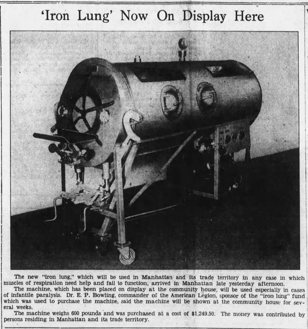
In September 1937, Charles Conroy Jr., a 17-year-old from Abilene with family in Manhattan, died of infantile paralysis in a Kansas City hospital, despite being placed in an iron lung. A week later the local post of the American Legion took the first steps to buy an iron lung for Manhattan.

The Legion and the Saint Mary hospital auxiliary asked the public to pay for it. Money came from the President’s Birthday Ball fund, from women’s clubs and lodges, from junior high students, from a dance at the college, a benefit movie at the Dickinson Theater and a wolf hunt. Gifts ranged from $1 to the $400 voted by the Mrs. Abraham Lincoln Tent No. 12.

By January 21, 1938, the fund stood at $1,084.22, and the goal of about $1,250 was soon reached.

The Manhattan Mercury, November 22, 1937.

A patient lies in a large tank respirator, an iron lung, with only the head outside the machine.

Manhattan Mercury page with a photograph of the new iron lung on display, April 12, 1938.

Ready at a Moment’s Notice

The iron lung went on display at the Community House, where townspeople came to see it. It was painted blue, and the Mercury thought it “quite attractive.” In November 1939 it moved to its permanent home at the fire station, to be used anywhere in the three-county area.

In August 1940 the health department had the police and firemen test it on three volunteers, one of them a Mercury reporter.

Breathing Easy in “Lung”

As the diaphragm moved in and out of the “lung,” it alternately compressed and relieved the air inside and the “patient” soon found it much more convenient to “breathe with the lung.” When the rubber mat was permitted to rest lightly against his throat thus making the tank almost air tight, the tester was able to relax and let the “lung” do his breathing for him.

At the end of a few minutes in the tank, this reporter decided he had slept on beds infinitely less comfortable than the “lung’s” and had to do his own breathing.

The Manhattan Mercury, August 26, 1940

Weeks later it was needed. On October 11, 1940, the iron lung was rushed to Fort Riley to treat a polio patient on the reservation, and it went back again in 1941 for a little girl at Camp Whitside. The flood of 1951 damaged it beyond repair.

Manhattan Mercury page with a photograph of the new iron lung on display, April 12, 1938.

Engraving of the respirator by Alfred F. Jones, 1864.

1864Plate 1 of 15

Alfred F. Jones

Lexington, Kentucky

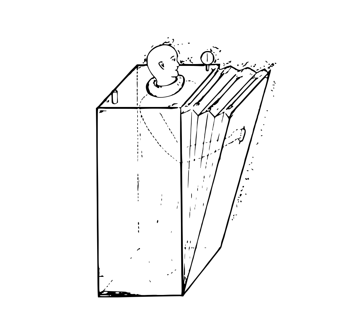
The earliest body-enclosing iron lung we know of was described by Alfred F. Jones of Lexington, Kentucky, in 1864. U.S. patent No. 44,198 says he “cured paralysis, neuralgia, rheumatism, seminal weakness, asthma, bronchitis, and dyspepsia. Also deafness… when properly and judiciously applied many other diseases may be cured…”

No. 1, Emerson exhibit

Engraving of the respirator by Dr. Woillez, 1876.

1876Plate 2 of 15

Dr. Woillez

Paris, France

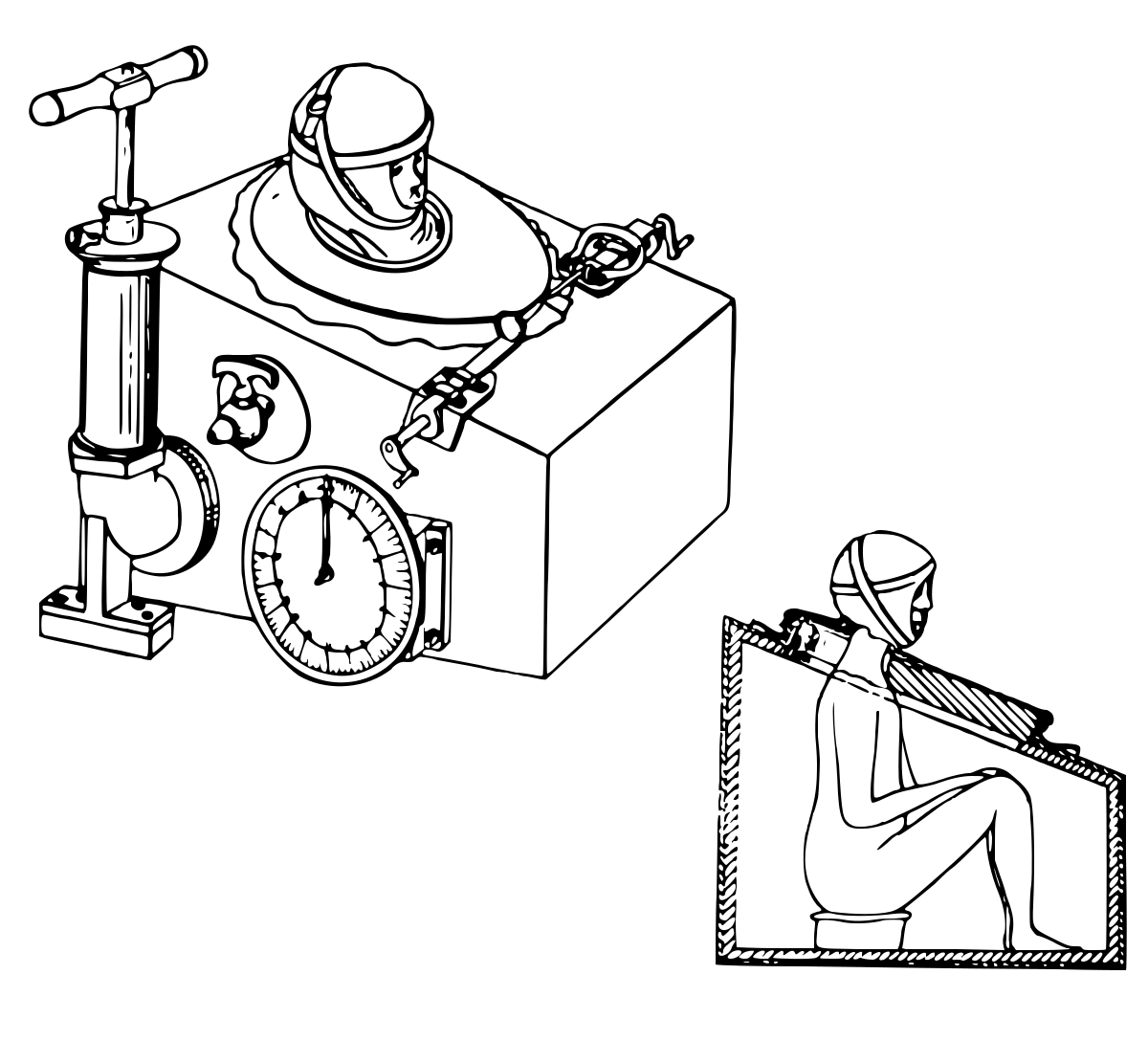
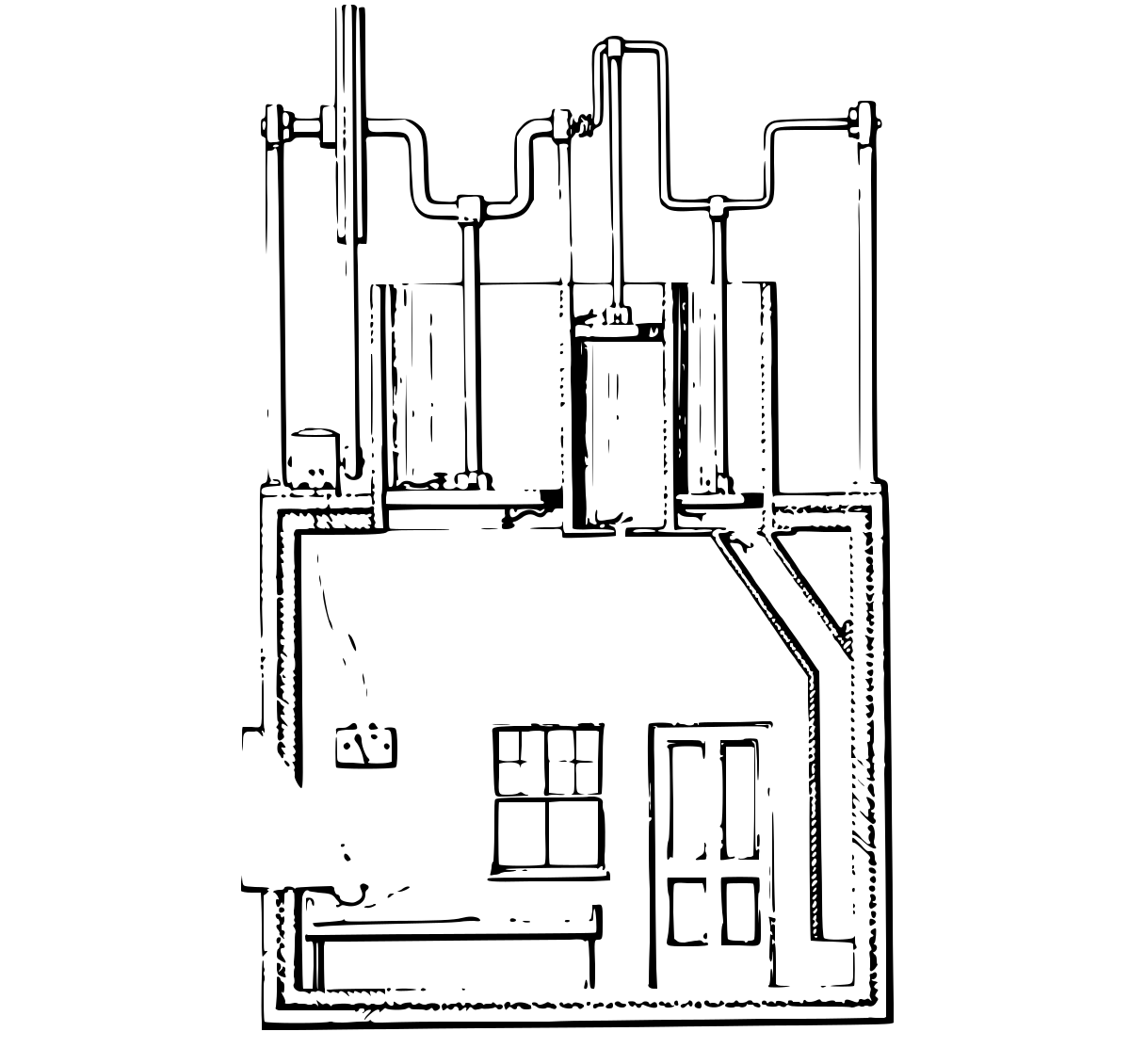
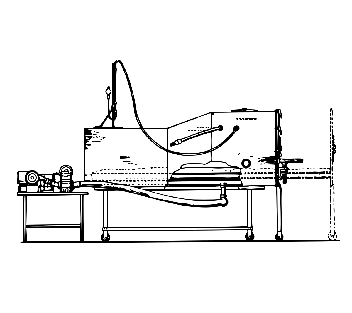
In 1876 Dr. Woillez of Paris built the first workable iron lung, which he called a “spirophore.” It had the basic elements of modern respirators, including an adjustable rubber collar and a sliding bed. A unique feature was a rod which rested lightly on the patient’s chest, to give visual proof of actual lung expansion. In a brilliant lecture before the Academy of Medicine on June 20, Woillez showed a thorough understanding of the physiology and mechanics of artificial respiration. He refused to patent his invention. A colleague suggested placing spirophores all along the Seine for drowning rescues, but the money for this public service was lacking. (The illustration was reconstructed by Maxfield Parrish, Jr., for a legal battle, years before the inset photograph was discovered.)

No. 2, Emerson exhibit

Engraving of the respirator by Dr. Charles Breuillard, 1887.

1887Plate 3 of 15

Dr. Charles Breuillard

Paris, France

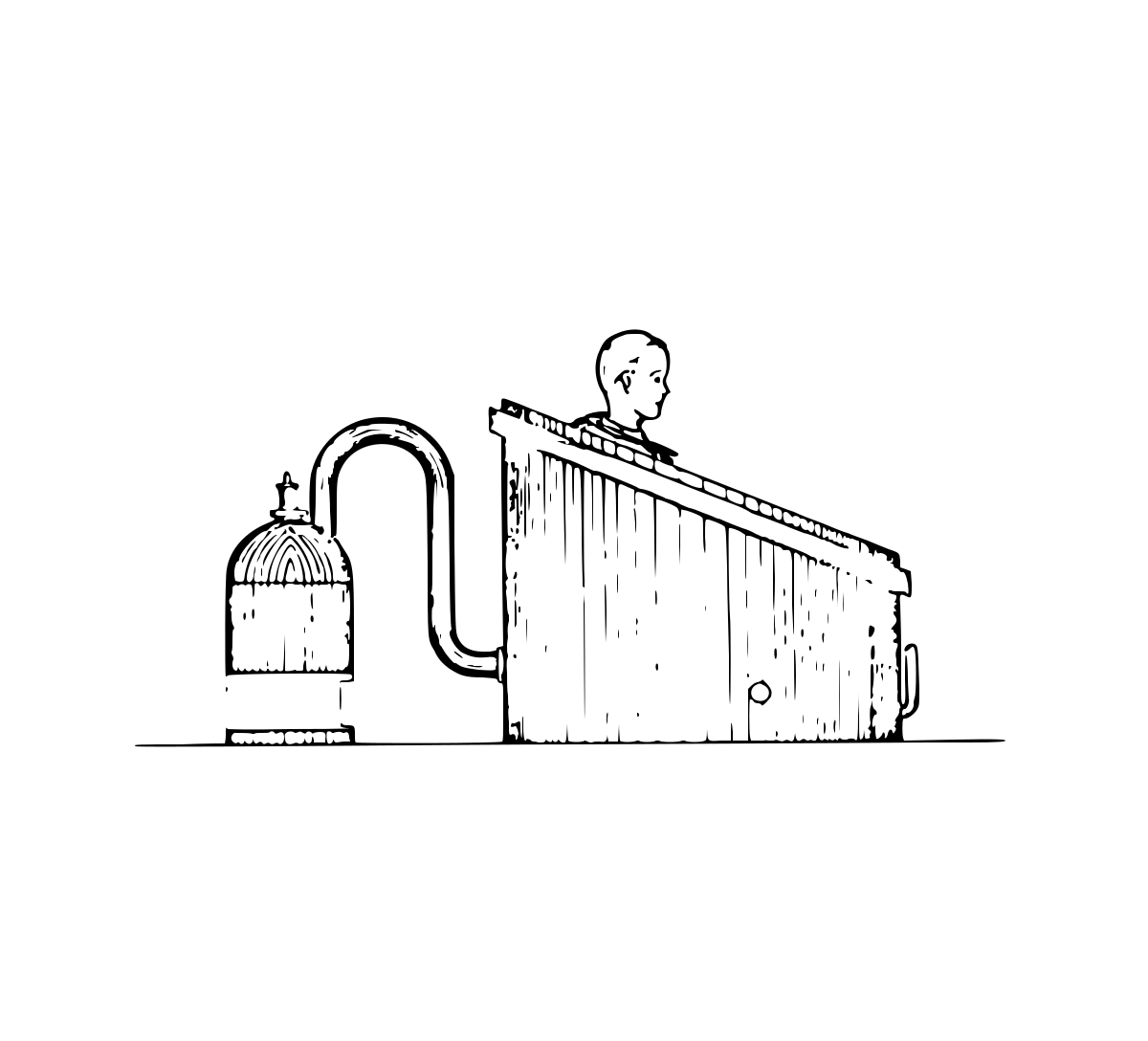
Dr. Charles Breuillard of Paris patented a “bath cabinet” type of respirator in 1887. For a source of vacuum he recommended “a steam ejector fed by a steam boiler… heated by a spirit lamp.” The patient himself was supposed to operate a valve, alternately connecting the cabinet with the vacuum for inhalation, and with the atmosphere for exhalation. Breuillard also described a chest respirator (“cuirass”) to be operated in the same way, and a face mask.

No. 3, Emerson exhibit

Engraving of the respirator by Dr. Egon Braun, 1889.

1889Plate 4 of 15

Dr. Egon Braun

Vienna, Austria

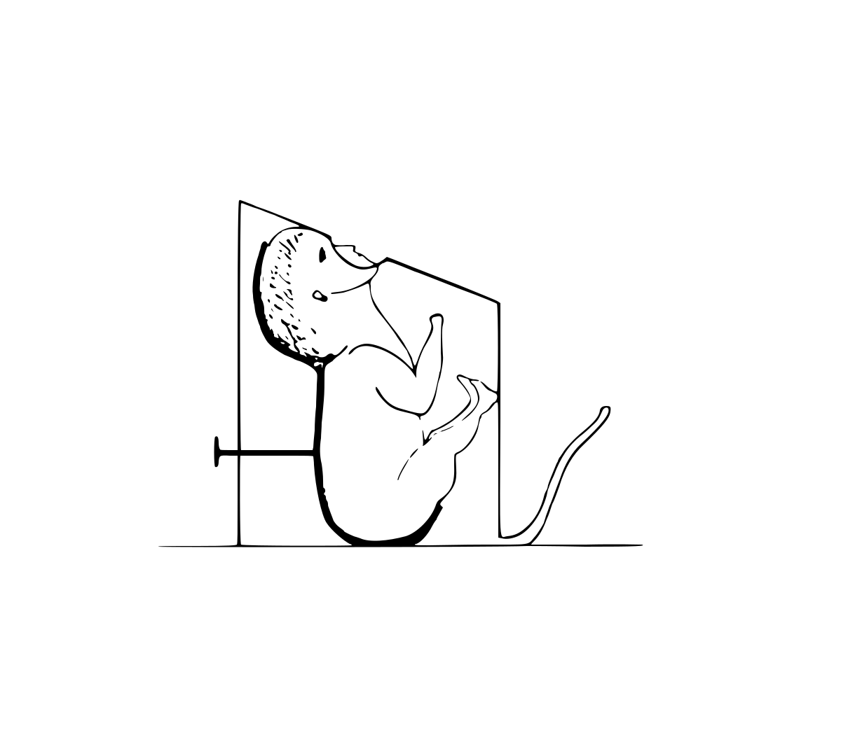
For “resuscitating asphyxiated children,” Dr. Egon Braun of Vienna devised a small wooden box in which pressure and suction were created by the doctor’s own breath (through the tube at the right). The infant was supported in a plaster mold, with nose and mouth pressed against an opening in the rubber closure. (This picture, from the Boston Medical and Surgical Journal of 1889, is only a rough diagram.) Dr. Braun reported 50 consecutive successful cases.

No. 4, Emerson exhibit

Engraving of the respirator by Rudolf Eisenmenger, 1901.

1901Plate 5 of 15

Rudolf Eisenmenger

Piski, Hungary

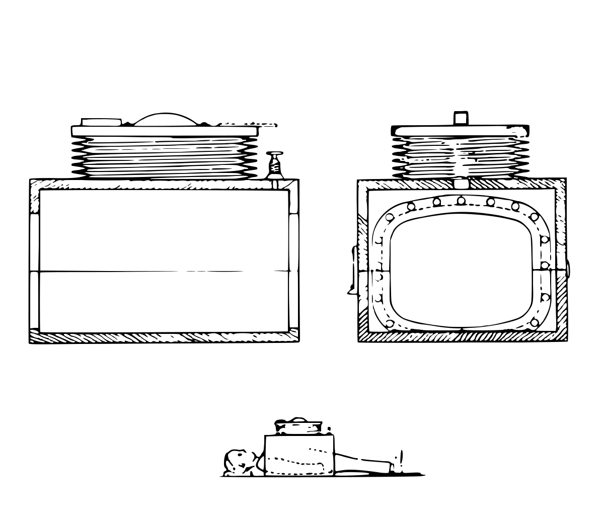
In 1901 Rudolf Eisenmenger of Piski, Hungary, patented a portable respirator which consisted of a “simple, two-part box” enclosing only the patient’s chest and abdomen. Later he became a medical professor in Vienna, and there continued to improve his invention. He stressed the importance of access to the patient’s throat and limbs, of portability, and of hand operation. (Motors were also mentioned.) There are reports of extraordinary success with Eisenmenger’s respirators.

No. 5, Emerson exhibit

Engraving of the respirator by William Davenport, 1905.

1905Plate 6 of 15

William Davenport

London, England

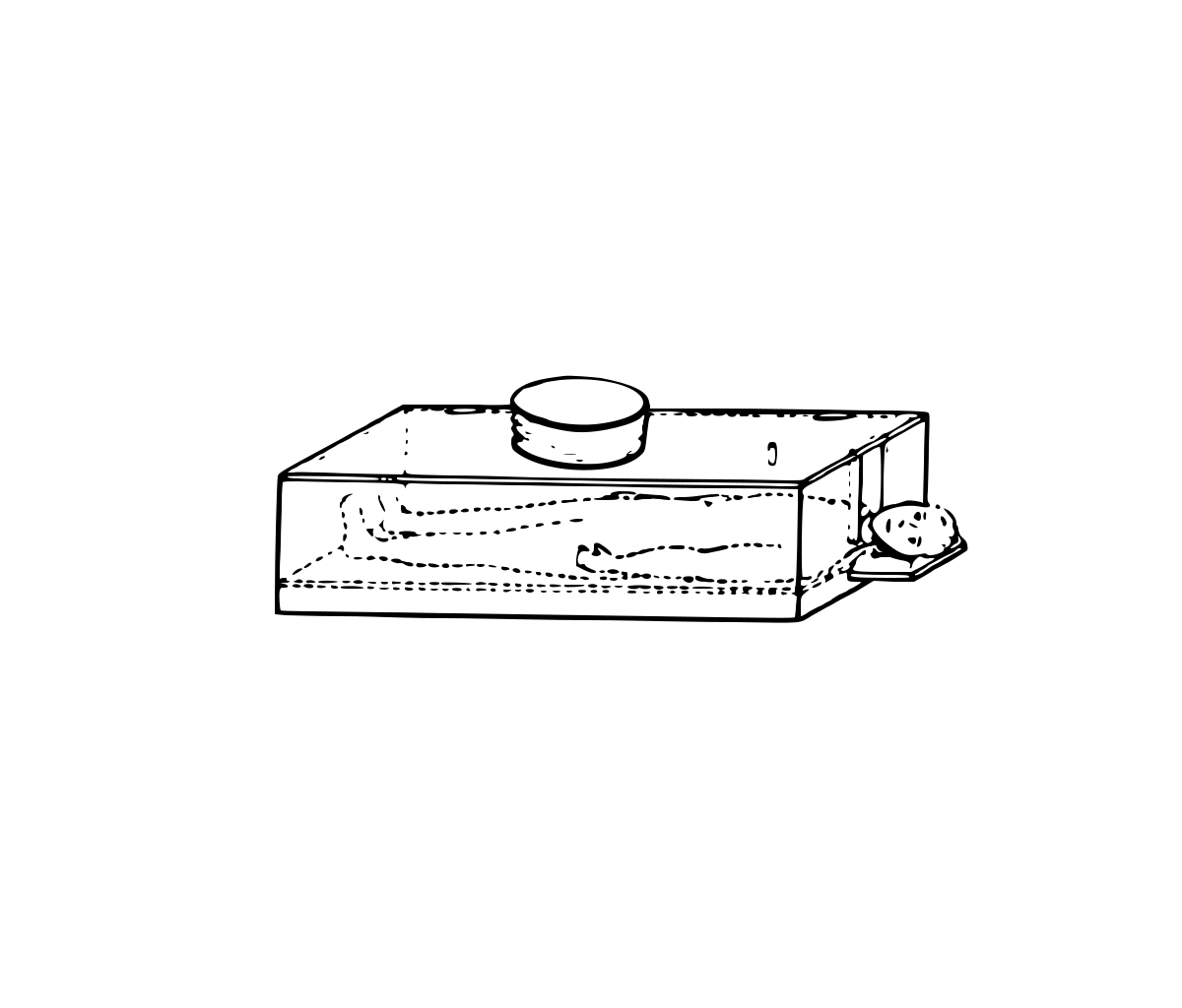
William Davenport of London understood the mechanics of artificial respiration clearly. His patent in 1905 mentions a box, a rubber collar, and a simple bellows or piston pump. He lacked the sliding bed (of Woillez and modern iron lungs) but made several good suggestions, including the supplementary use of oxygen. He proposed several types, including a “collapsible form… to facilitate transit.”

No. 6, Emerson exhibit

Engraving of the respirator by Dr. Charles Morgan Hammond, 1905.

1905Plate 7 of 15

Dr. Charles Morgan Hammond

Memphis, Tennessee

Dr. Charles Morgan Hammond of Memphis built his first “artificial lung” in 1905, and a series of improved models over the next 20 years. He performed experiments and treated patients successfully, but his respirators were not produced commercially and remained unknown to the public for many years.

No. 7, Emerson exhibit

Engraving of the respirator by Peter Lord, 1908.

1908Plate 8 of 15

Peter Lord

Worcester, Massachusetts

In 1908 Peter Lord of Worcester patented a respirator room. He pictured huge pistons to create the pressure changes and to supply fresh air. An enormous motor would have been needed, and very heavy construction. However, the idea of a room is good, because it makes nursing care easy.

No. 8, Emerson exhibit

Engraving of the respirator by Melvin L. Severy, 1916.

1916Plate 9 of 15

Melvin L. Severy

Boston, Massachusetts

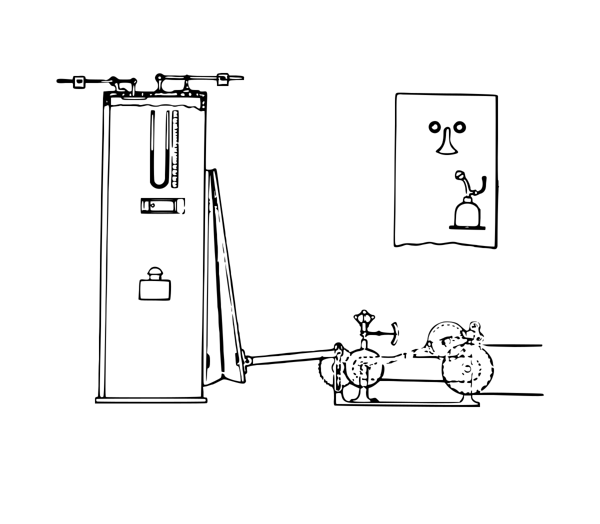
The patent granted to Melvin L. Severy of Boston in 1918 shows little concern for the patient’s comfort but includes ingenious mechanical elements. In the model illustrated, the patient was obliged to stand, pressing his nose and mouth against a triangular opening below two eye windows. Severy also described variable-speed pulleys and elaborate electromagnetic controls for regulating speeds and pressures. He suggested cams to produce various pressure curves. A cuirass respirator and mask resuscitator were mentioned also.

No. 9, Emerson exhibit

Engraving of the respirator by Dr. W. Steuart, 1918.

1918Plate 10 of 15

Dr. W. Steuart

South Africa

Dr. W. Steuart of South Africa built a simple respirator and demonstrated a working model to his colleagues (Medical Journal of South Africa, 1918). He proposed plaster of Paris to seal around the patient’s shoulders and pelvis, but friends quickly suggested more practical materials. Steuart was the first to think primarily of poliomyelitis. He even imagined a ward piped with intermittent suction, to which individual body casings could be attached.

No. 10, Emerson exhibit

Engraving of the respirator by Chillingworth and Hopkins, 1919.

1919Plate 11 of 15

Chillingworth and Hopkins

New Orleans, Louisiana

Felix P. Chillingworth and Ralph Hopkins of New Orleans built a plethysmograph to study the circulatory changes caused by lung distension. They also used it as a dog respirator. “…pulmonary ventilation is accomplished by rhythmic changes in air pressure within the plethysmograph causing alternate distention and collapse of the lungs.” (Journal of Laboratory and Clinical Medicine, 1919.)

No. 11, Emerson exhibit

Engraving of the respirator by Wilhelm Schwake, 1926.

1926Plate 12 of 15

Wilhelm Schwake

Oranienburg-Eden, Germany

Wilhelm Schwake of Oranienburg-Eden, Germany, patented a “pneumatic chamber” in 1926. He was concerned with portability and with exactly matching the patient’s depth and rhythm of breathing. He also stated that “negative pressure… upon the skin… draws out… the gaseous by-products.” We would not consider this respirator practical for severely ill patients, but the picture is a great favorite.

No. 12, Emerson exhibit

Engraving of the respirator by Philip Drinker and Louis Agassiz Shaw, 1928.

1928Plate 13 of 15

Philip Drinker and Louis Agassiz Shaw

Boston, Massachusetts

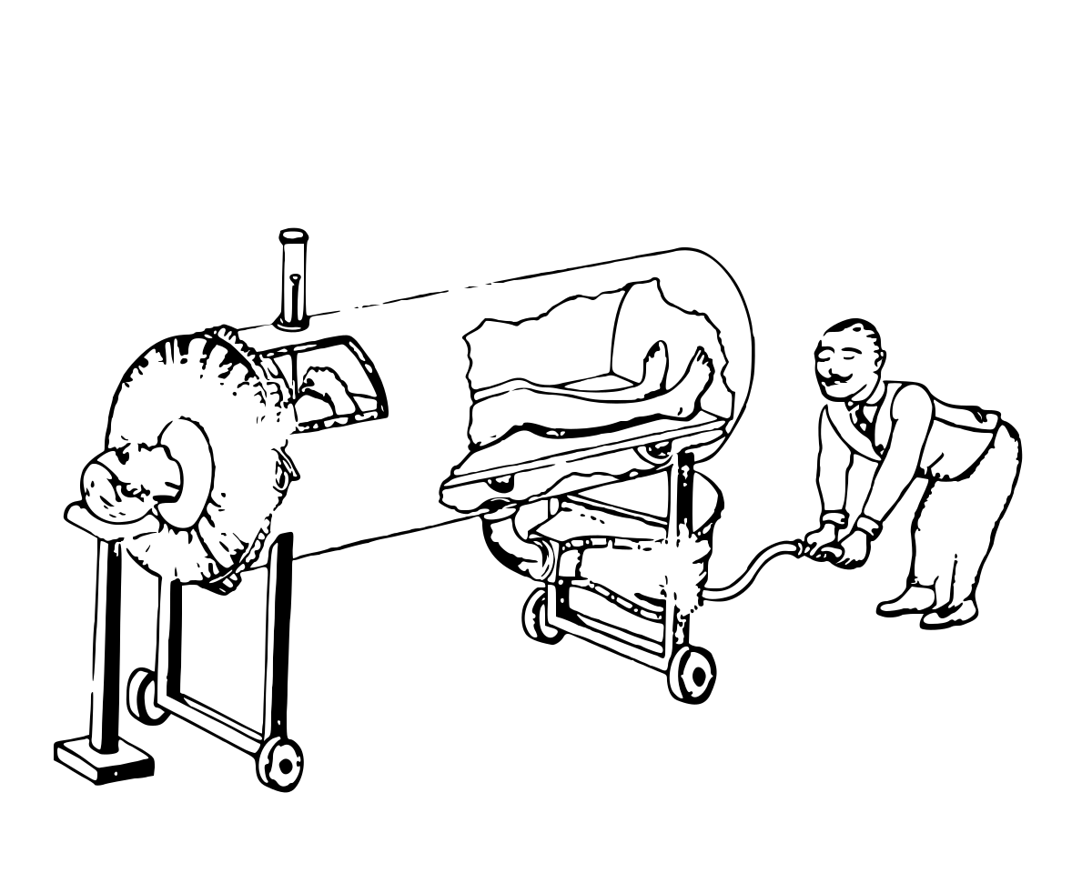
The first “iron lung” to receive widespread use was developed in 1928 in Boston, and was patented by Philip Drinker and Dr. Louis Agassiz Shaw. It was cumbersome and inconvenient but saved a number of lives. It had a sliding bed with a headwall attached and a rubber collar. Pressure changes were created by a rotary blower and an alternating valve. During a polio epidemic the Consolidated Gas Company of New York paid for building large numbers of these respirators, and reports of their use spread quickly around the world.

No. 13, Emerson exhibit

Plate 14: Wilson and Drinker.

UndatedPlate 14 of 15

Wilson and Drinker

Boston, Massachusetts

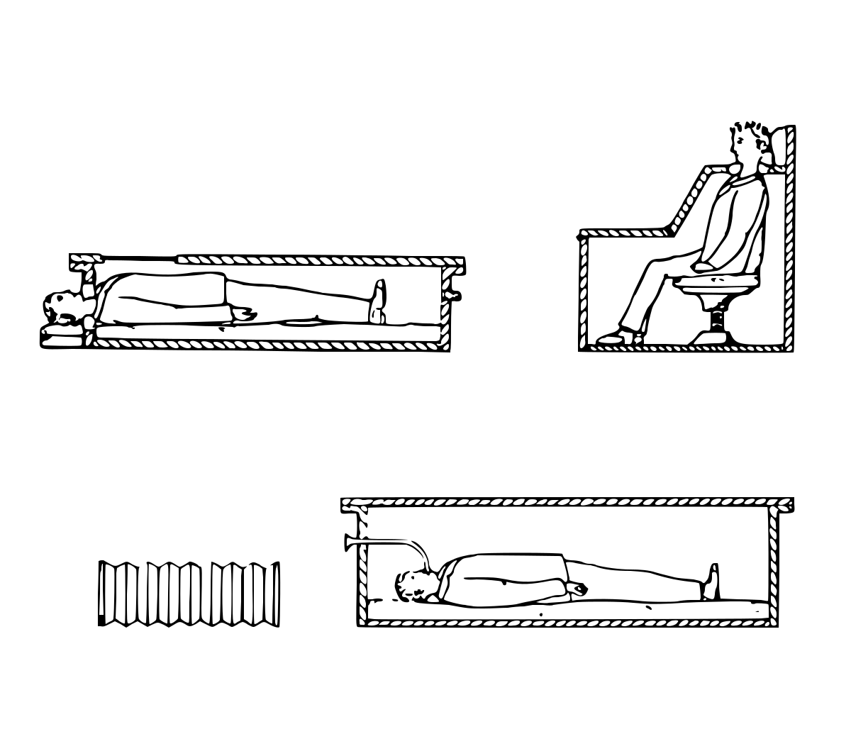
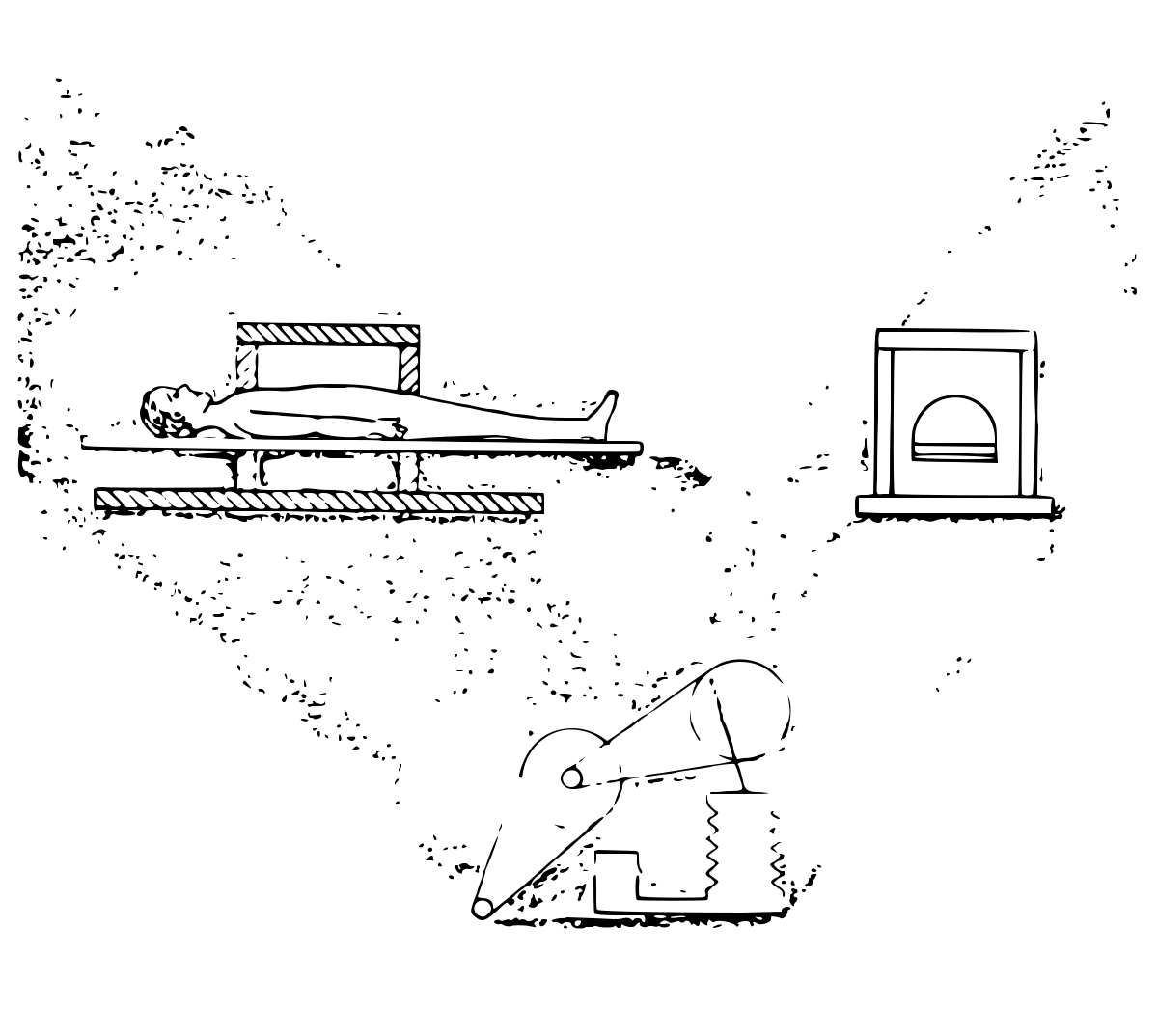
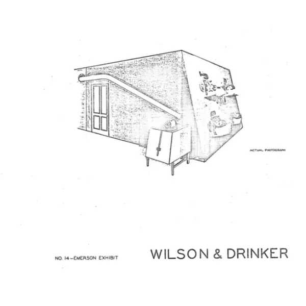
In the early respirators it was difficult to give complete nursing care and to change the patients frequently. This care was found to be of life-saving importance, so Dr. James L. Wilson asked for a room in which several patients could be made to breathe at the same time. A nurse could enter by the door and perform all procedures efficiently. The room pictured was built at the Children’s Hospital in Boston and was used successfully during several epidemics.

No. 14, Emerson exhibit

Plate 15: John Haven Emerson.

1931Plate 15 of 15

John Haven Emerson

Cambridge, Massachusetts

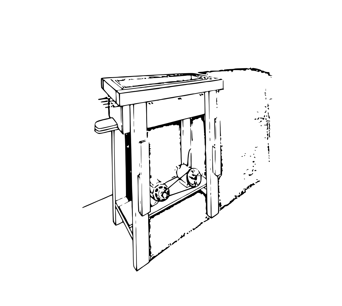
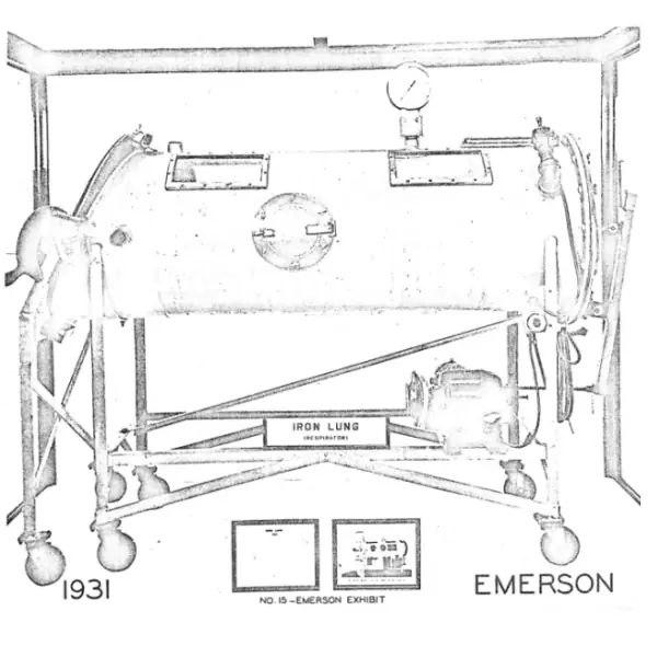
Because of a severe poliomyelitis epidemic in 1931, John Haven Emerson of Cambridge built a simplified respirator. It cost less than half as much as others, but contained many improvements. It operated quietly, using a bellows to create the changes of pressure (as Woillez’s did, but with a motor added). A wide range of speeds was instantly available. Opening and closing were rapid and convenient. It could be pumped by hand if the electricity failed. The first Emerson “iron lung” is now preserved at the Smithsonian Institution as the prototype of the respirators built since 1931 in America and Europe. Manhattan bought one of its machines in 1938.

No. 15, Emerson exhibit

Iron Lung, Iron Will

From the Mercury

The iron lung fund drive of 1937 and 1938, and the polio epidemic that closed Manhattan’s schools in September 1940, as Manhattan’s newspapers reported them. Tap an entry to read the article and see the clipping.

  1. Loading the timeline…
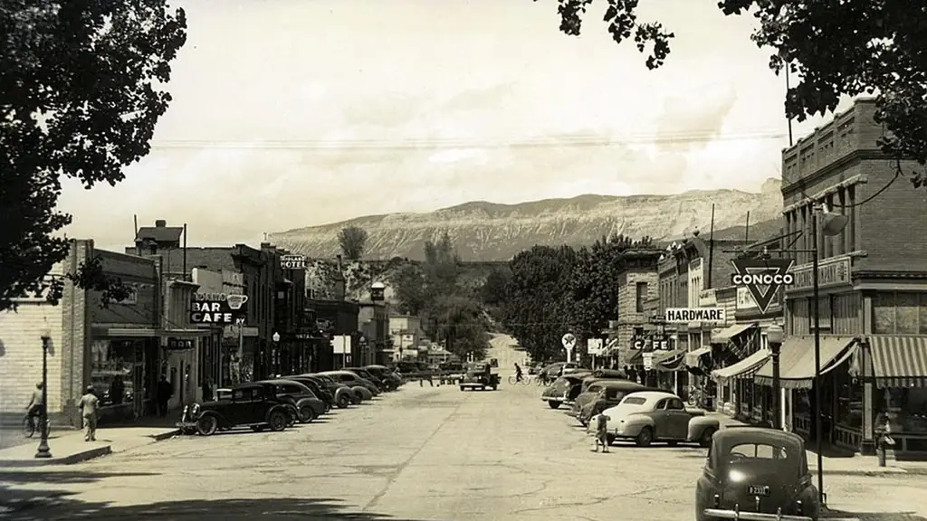
A small-town main street lined with shops and parked cars, mid-twentieth century.

Policemen were stationed at every entrance to the city [of Hoboken, New Jersey]—tube, train, ferry road, and cow path—with instructions to turn back every van, cart, and person…

New York Times, July 14, 1916

Communities

Polio occurred primarily in July, August, and September and hit regardless of geographic region, economic status, or population density. Relatively few people showed any symptoms and even fewer died or experienced paralysis, but the physical effects were dramatic. Communities reacted with dread because no one understood how or why people got it, and because children were the most frequently affected.

Sign barring children under sixteen from entering town, posted on a tree during the New York City epidemic, 1916.

Many inspectors… stationed themselves at the railroad stations, ferries, and boat landings along the Delaware River… to bar all children under 16 years of age who attempted to cross into [Pennsylvania] without certificates of health.

Los Angeles Times, August 9, 1916

A small-town main street lined with shops and parked cars, mid-twentieth century.

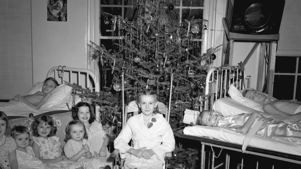
Children in hospital beds and wheelchairs gathered around a Christmas tree in a polio ward.

In isolation, it ripped my body from me, without warning. There was no possible preparation, or forethought. I woke up after the searing jumble of fever as an eighteen-year-old boy in a seventy-five-year-old body.

Lorenzo Wilson, 1984

Families and Individuals

The enforced separation of families during the early, acute phase of the disease contributed to the intense dread and fear that polio aroused. Children and parents were not allowed any contact for ten to fourteen days and then only limited visiting for weeks afterward. When the person returned home weeks or months later, adjustment to changed circumstances brought more stress.

I spent my first 16 years in hospitals. Since I was in a place that was a twelve-hour drive away from my family, I rarely got visited. Every Sunday was visiting hours. Each girl had to get dressed, bed made, and quietly sit for the duration of the visiting hours. You had to make a decision to either be sad that you didn’t have any company, or be happy for those who did.

Georgia Gibson, 2004

Children in hospital beds and wheelchairs gathered around a Christmas tree in a polio ward.

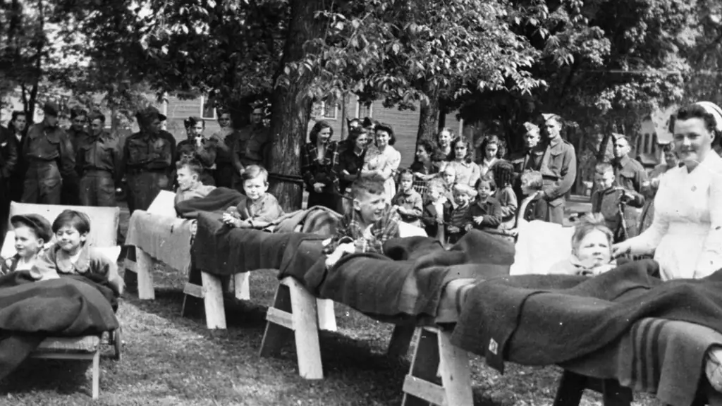
Rows of children lying in makeshift beds outdoors, watched by soldiers, nurses and families.

In no disease, except perhaps pneumonia, is expert nursing care so essential.

Dr. T. Campbell Thompson, 1938

The Medical Community

Throughout most of the 20th century, hospitals operated under strict and orderly patient regimes. Nurses enforced a military-like discipline in the wards. Epidemic conditions, combined with the lack of a cure for polio, heightened everyone’s anxiety.

During a 1934 epidemic in Los Angeles, 5 percent of doctors and 11 percent of nurses who treated polio patients contracted the disease.

In the town of Hickory, in the western foothills of North Carolina, more than 100 children lie in hastily built beds… The suddenness with which the disease struck overwhelmed ordinary medical facilities… The National Foundation for Infantile Paralysis mobilized local and national resources to set up the emergency hospital.

LIFE magazine, 1944

Rows of children lying in makeshift beds outdoors, watched by soldiers, nurses and families.

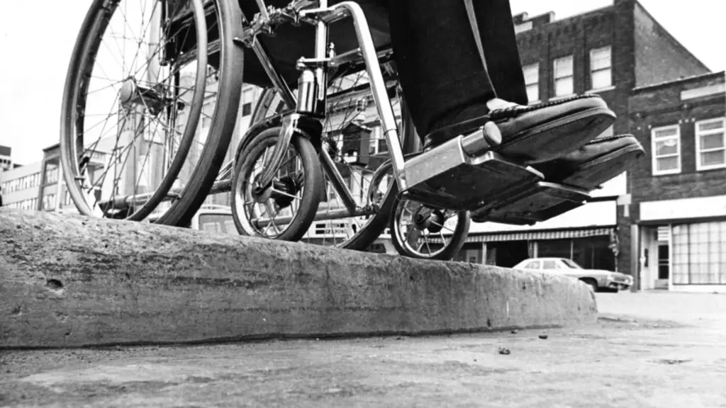
A wheelchair stopped at the edge of a high street curb.

The systemic denial of the chance to work, joined on education, marriage, and most forms of social intercourse, have entailed what amounts to… social death.

Paul Longmore, 2003

Disability Rights

Many people who had been injured by the poliovirus became discouraged when they returned home and encountered environmental barriers and discrimination. Some were among the most eloquent and influential leaders of the disability rights movement. Their message has been that disability is a social and civil rights issue, not simply a medical problem.

After a series of incremental policy revisions to aid people with disabilities, in 1990, the Americans with Disabilities Act was passed by the U.S. Congress. The bill became landmark civil rights legislation which creates broad legal protections for people with disabilities.

In 1968, Hugh Gallagher and [Alaska Senator Bob] Bartlett cooked up the Architectural Barriers Act which stated that all buildings ‘designed, built, altered, or leased with Federal funds’ would be required to have ramps, curb cuts and access to all facilities. It was their radical view that accessibility was a basic civil right for all.

Lorenzo Wilson Milam, 2004

A wheelchair stopped at the edge of a high street curb.

Let no person ever again be segregated in an institution, a back room or any other place for lack of services or communities that empower.

Justin Dart, 1998

Social Effects

The effects of polio can be found throughout American culture: in the lives of people who survived it, through changes to philanthropy, and in new approaches to the design of everyday objects.

Frank C. Mars, seen here in the 1890s, contracted polio as an infant. Mars Milky Way candy box from around 1930 and Mars company candy package.

My father, Frank C. Mars, had polio as a baby and he couldn’t walk until he was around twelve years old. The nearest school was a number of miles away and he couldn’t walk there. As a consequence, his mother Elva home-schooled him. This meant long hours in the kitchen where Elva prepared meals for the family and taught Frank his lessons. Elva was a capable candy maker and hand-dipped chocolate for her two sons.

Forrest Mars Sr., 2005
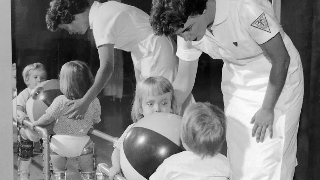
Nurses helping young children in leg braces exercise with a large ball.

The goal of total rehabilitation is to teach the physically handicapped person to live not just within the limits of his disability but to live to the hilt of his capabilities.

Dr. Howard Rusk, 1946

The Medical World

Polio patients were most vulnerable in the acute stage, when the virus was actively destroying the motor neurons that controlled the muscles connected to swallowing, breathing, and limb movement. Although there was, and is, no cure for polio, endangered lives could be saved. Doctors and nurses used technology, experience, and vigilance to keep patients alive until the infection ran its course, and recovery began.

Sister Kenny (right) applying hot packs in 1943.

Elizabeth Kenny, or Sister Kenny, as nurses were called in Australia, came to the United States in 1940. Her methods of hot-pack applications, stretching, and muscle massage were unconventional and controversial, but eventually became part of standard care for polio.

The scientific community and the government learned valuable lessons in response to polio. There were changes in government oversight of vaccine development and surveillance of clinical trials for vaccines and drugs, in design of laboratory facilities, and in the nature of rehabilitation.

Nurses helping young children in leg braces exercise with a large ball.

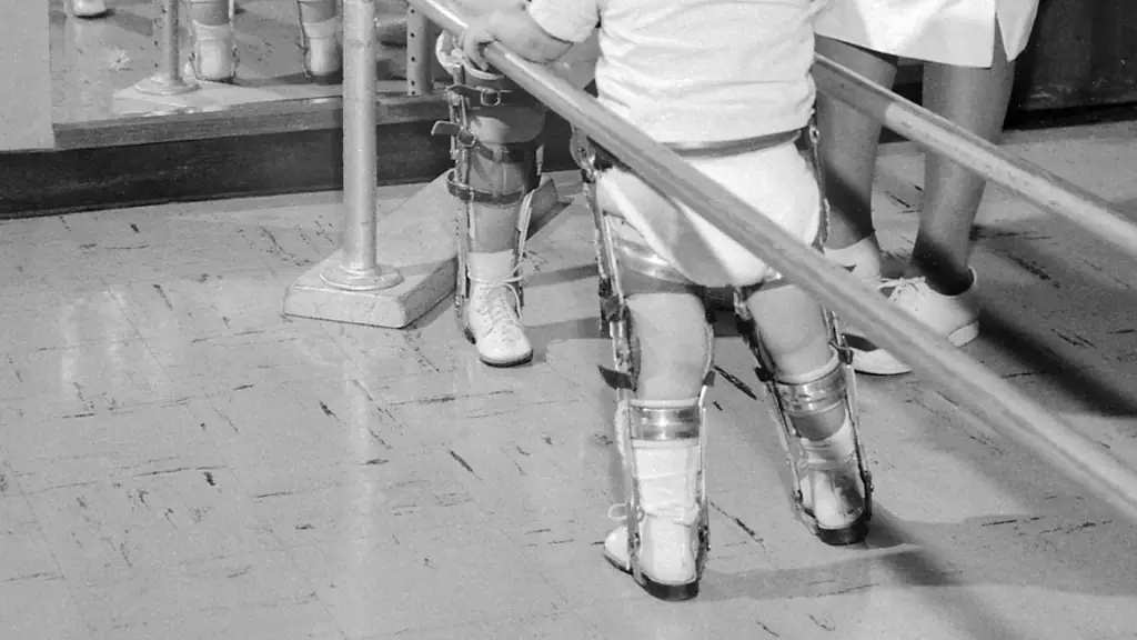
A child in steel leg braces and leather cuffs walking between parallel bars.

Toward Rehabilitation

A child in steel leg braces and leather cuffs walking between parallel bars.

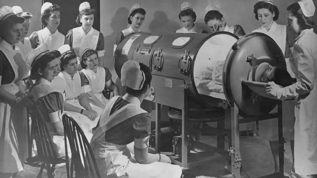
Nurses gathered around a row of iron lung respirators, a patient’s head visible at the end of one tank.

The Iron Lung

Nurses gathered around a row of iron lung respirators, a patient’s head visible at the end of one tank.

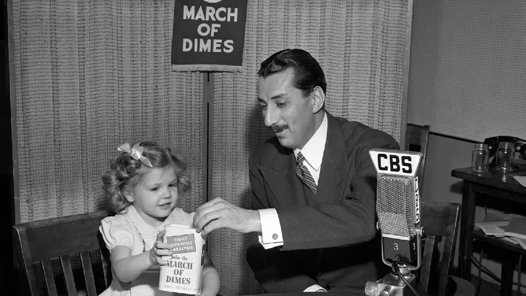
A young girl drops a coin into a March of Dimes collection box held by a man at a CBS radio microphone.

I couldn’t believe it, and I made [my wife] go over it again and again. The [March of Dimes] had been set up for cases like mine, people who couldn’t possibly meet the overwhelming expenses involved in an illness like this, or who, by meeting it, would find themselves hopelessly in debt.

Larry Alexander, 1954

Medical Philanthropy

Three private organizations figured prominently in the history of poliomyelitis in the United States and worldwide: the National Foundation for Infantile Paralysis (March of Dimes), the Rockefeller Institute, and Rotary International.

In answering a question regarding who would own the patent to his recent vaccine:

Well, the people, I would say. There is no patent. Could you patent the sun?

Jonas Salk, 1955

A young girl drops a coin into a March of Dimes collection box held by a man at a CBS radio microphone.

A young girl drops a coin into a March of Dimes collection box held by a man at a CBS radio microphone.

The March of Dimes

A young girl drops a coin into a March of Dimes collection box held by a man at a CBS radio microphone.

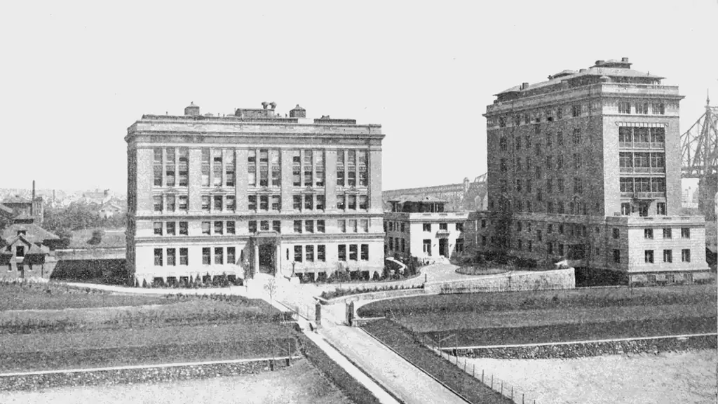
The buildings of the Rockefeller Institute for Medical Research in New York, early twentieth century.

Rockefeller University

The buildings of the Rockefeller Institute for Medical Research in New York, early twentieth century.

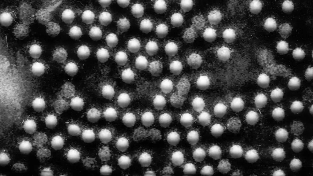
A magnified field of round virus particles.

To initiate infection, [the] virus must first attach to its cell-surface receptor and then deliver its RNA to the appropriate cellular compartment.

David Belnap, biochemist, 2000

How the Poliovirus Works

Poliomyelitis is caused by a poliovirus. Viruses are particles of either RNA or DNA and are neither living nor dead. They cannot do anything until they find an appropriate cell whose internal mechanism they can commandeer to start reproducing more replicas of their viral proteins.

There are three types of poliovirus and many strains of each type. The virus enters through the mouth and multiplies in the throat and gastrointestinal tract, then moves into the bloodstream and is carried to the central nervous system where it replicates and destroys the motor neuron cells. Motor neurons control the muscles for swallowing, circulation, respiration, and the trunk, arms, and legs.

Human nerve cells have a protruding protein structure on their surface whose precise function is unknown. When poliovirus encounters the nerve cells, the protruding receptors attach to the virus particle, and infection begins. Once inside the cell, the virus hijacks the cell’s assembly process and makes thousands of copies of itself in hours. The virus kills the cell and then spreads to infect other cells.

A magnified field of round virus particles.

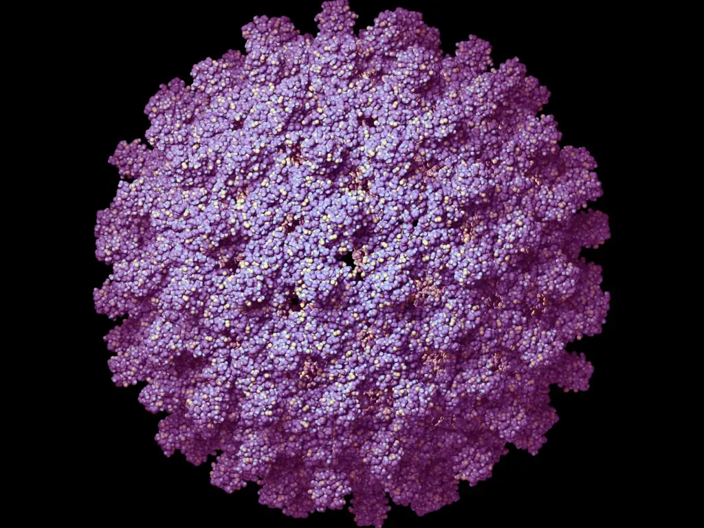
Cut-away illustration of a poliovirus particle, about 30 nanometres across. Its shell is built from 60 copies each of the proteins VP1 (blue), VP2 (green) and VP3 (orange), enclosing a single strand of RNA (gold).

Virology

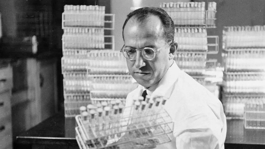
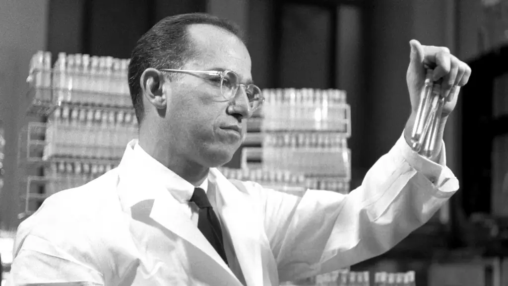
Jonas Salk in a white lab coat holding a rack of test tubes in front of shelves of glassware.

Chief objective: To determine whether a… poliomyelitis vaccine would afford protection against naturally occurring paralytic poliomyelitis in selected groups… and if protection is provided, the degree of such protection.

Evaluation of the 1954 Field Trial of Poliomyelitis Vaccine: Final Report, 1957

The Salk Vaccine

From the early 1900s, researchers pursued two different kinds of polio vaccine. One used inactivated (killed) viruses. The other kind used live but attenuated, or weakened, virus. The leading proponent of the killed virus was Jonas Salk.

The chief advantage of Salk’s killed virus vaccine was safety. If made properly, it could not cause disease. Its chief disadvantage was that the formaldehyde used in its manufacturing process caused the immune system to recognize killed virus differently from the live virus, possibly risking a shortened period of immunity.

While in his second year at New York University Medical school, Jonas Salk heard two lectures that changed his life: ‘In the first lecture, we were told that it was possible to immunize against diphtheria and tetanus by the use of a chemically treated toxin [to kill it]… In the very next lecture, we were told that in order to immunize against a virus disease, it was necessary to go through the experience of infection. It was not possible to kill the virus… The light went on at that point. I said that those two statements can’t possibly both be true. One has to be false.’

Jonas Salk, 1990

Jonas Salk in a white lab coat holding a rack of test tubes in front of shelves of glassware.

Jonas Salk holding up a test tube and examining it, racks of glassware behind him.

Polio Pioneers

Jonas Salk holding up a test tube and examining it, racks of glassware behind him.

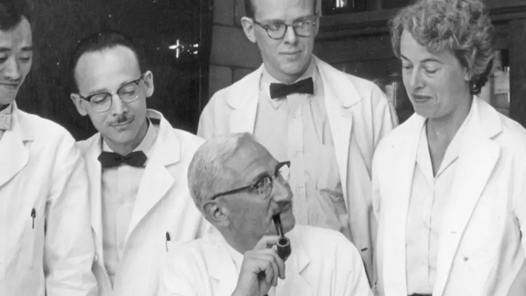
Albert Sabin with a pipe, surrounded by colleagues in lab coats.

I have studied the effects of our new lots of polio vaccine in 100 adult volunteers and during the next few days shall give it to my wife and 2 children as well as to our neighbors and their children.

Albert Sabin, 1957

The Sabin Vaccine

While the large-scale clinical trial with Salk vaccine went ahead in 1954, Albert Sabin pursued development of a live-virus vaccine. Like many researchers of the day, Sabin strongly disagreed with Salk’s approach of using injected, “killed” virus. He believed that long-term immunity could only be achieved with a live, attenuated—or weakened—virus.

Sabin and other researchers had argued that long-term immunity to polio could only be achieved with a live though greatly weakened virus, and that it must follow the same route of infection, through intestinal tissue. Weakening the virus required passing it through a succession of animals: rats, mice, or monkeys. This allowed it to become more virulent for these hosts, and less so for humans.

An important feature of Sabin’s oral polio vaccine was that immediately after vaccination, people shed weakened virus in their fecal waste. This boosted immunity for others in the community and gradually reduced the number of people susceptible to poliomyelitis.

Albert Sabin with a pipe, surrounded by colleagues in lab coats.

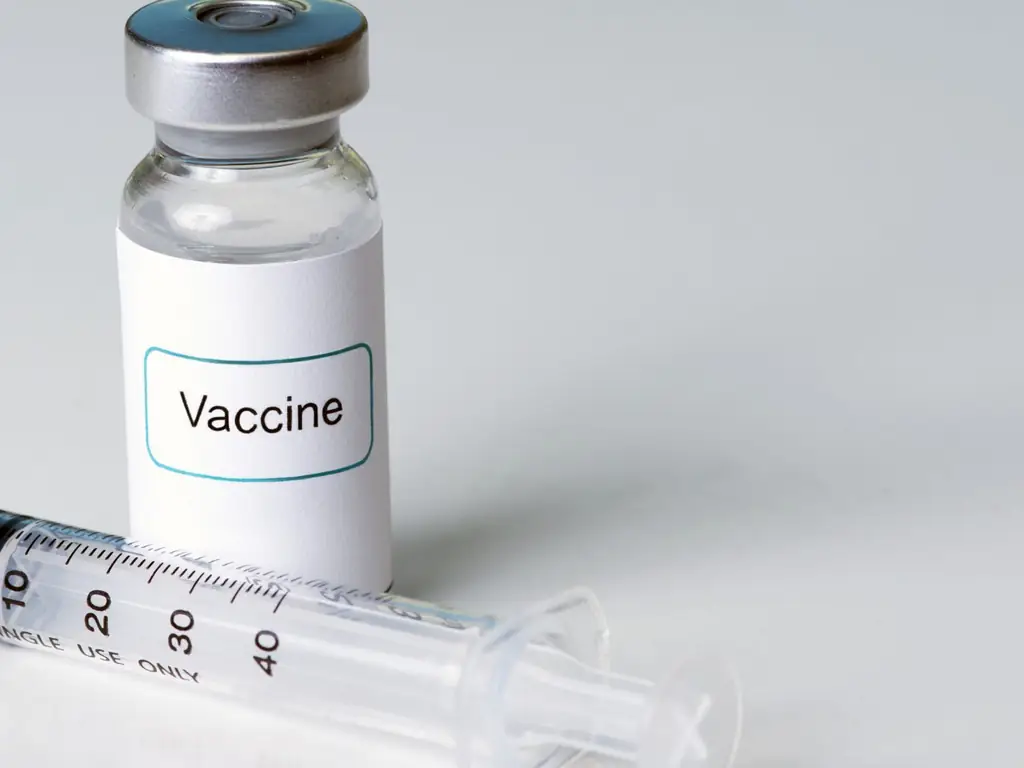
A small glass vial labelled “Vaccine” beside a syringe.

Introduction

Vaccines have greatly reduced the prevalence of disease, and they continue to be important for global health today. This part of the exhibit explains how vaccines work, which diseases they prevent, and who in the world is protected by them.

There is a technical difference between vaccination and immunization, as Britain’s National Health Service explains:

Vaccination means having a vaccine, that is actually getting the injection, or nasal spray or oral vaccine. Immunization means both receiving a vaccine and then becoming immune to a disease.

National Health Service (NHS)

The distinction is made because in a very small number of people the vaccination will not “take,” and that person will not be immune to the disease. Because this is so rare, the two terms are often used interchangeably.

A small glass vial labelled “Vaccine” beside a syringe.

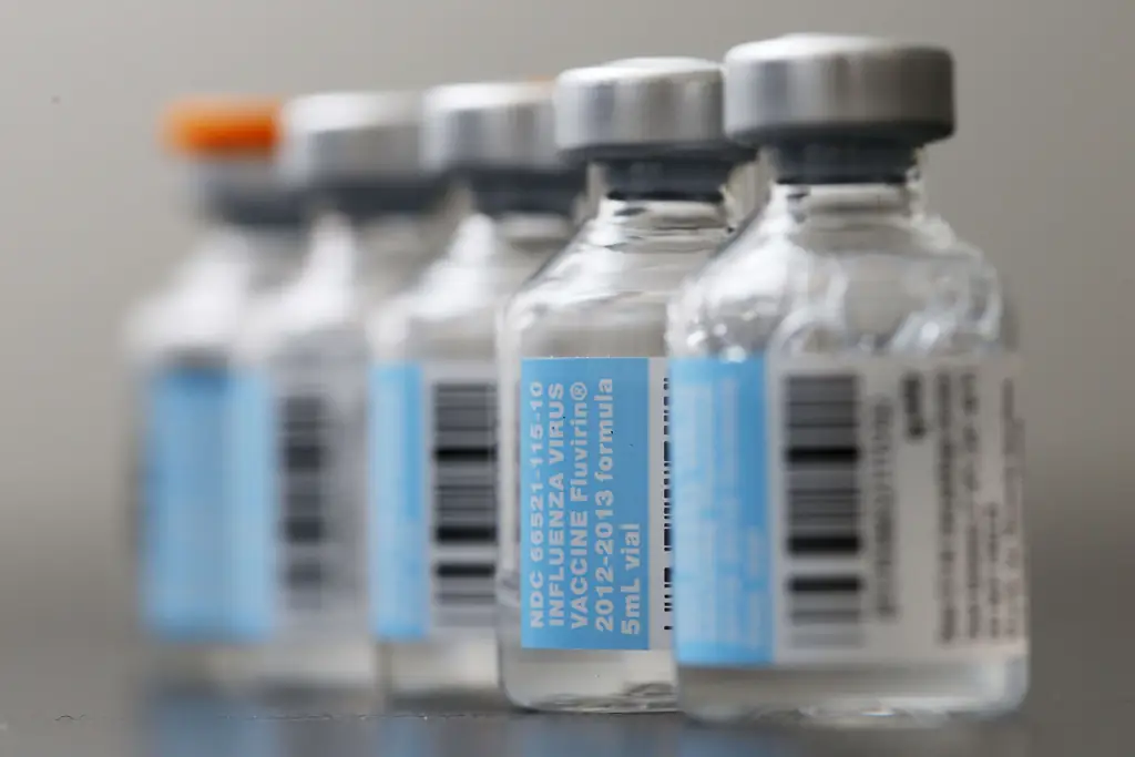
A row of small vaccine vials with blue labels, out of focus.

How Vaccines Work

A vaccine trains the immune system to recognize a germ before the real infection ever arrives.

How a vaccine works
  1. 1

    The vaccine brings a killed or weakened form of the virus or bacteria into the body.

  2. 2

    The immune system makes antibodies to defend against it.

  3. 3

    When the real, active germ enters the body later, the defense system recognizes it and can eliminate it.

  4. 4

    Vaccines strengthen the body’s natural defenses without causing the illness.

Because the body has already made antibodies and memory cells, it can fight off the real virus or bacteria quickly, often before a person ever feels sick.

A row of small vaccine vials with blue labels, out of focus.

A painting of a Chinese physician blowing powdered smallpox scabs into a child’s nose through a long tube.

The Origin of Vaccination

Vaccination has a long history. An early form was called variolation, or more broadly inoculation. Practised for centuries in Asia, it was a technique of deliberate smallpox infection: dried smallpox scabs were blown up the nose to give the person a form of the disease that was often milder.

By the 1700s variolation had spread to Africa, India and the Ottoman Empire, and then to Britain and America, where the infection was more often given through a puncture in the skin.

Variolation did work, but the risks were large. People who were variolated could catch the severe form of smallpox and die, and they could also pass the disease on to others.

A painting of a Chinese physician blowing powdered smallpox scabs into a child’s nose through a long tube.

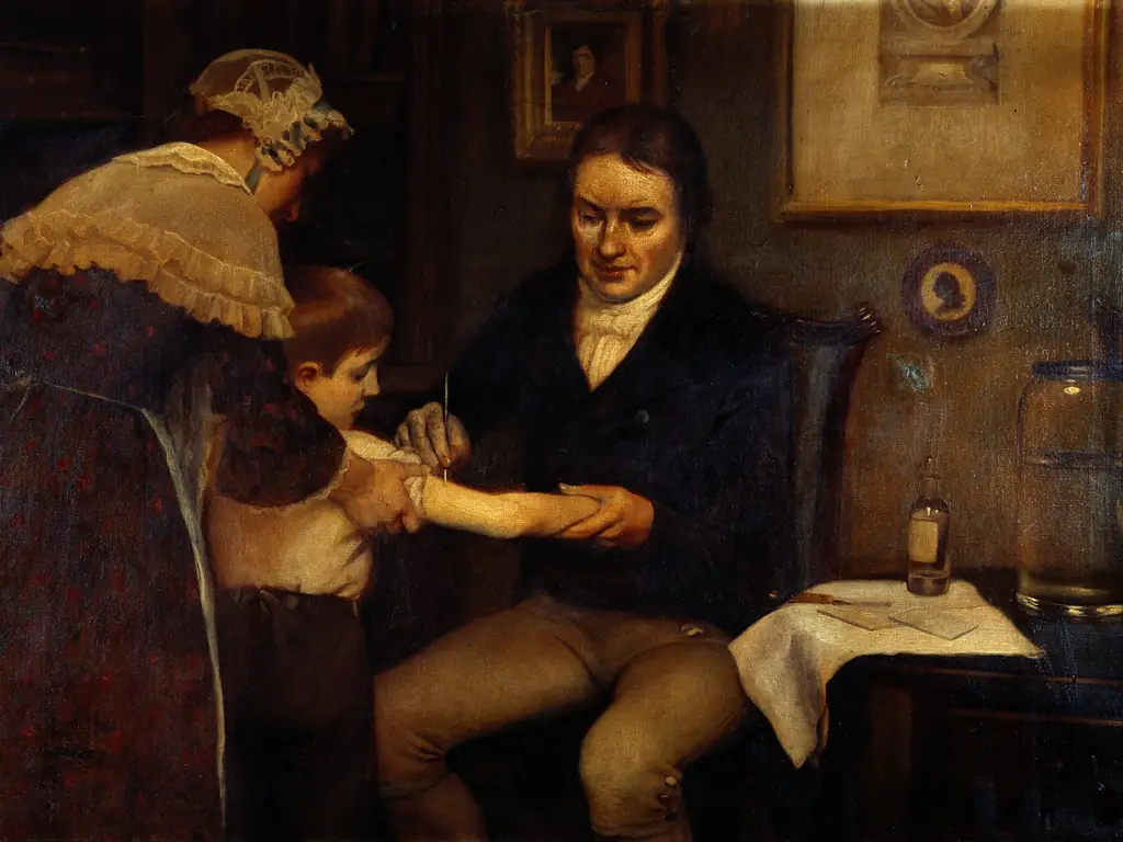
A painting of Edward Jenner vaccinating a young boy as a woman holds the child.

A Scientific Method

In 1796 the English physician Edward Jenner demonstrated another method of inoculation, using cowpox. Cowpox is a disease similar to smallpox, and it had been observed that a cowpox infection could protect against smallpox.

Jenner took matter from a cowpox sore and inoculated his gardener’s eight-year-old son, James Phipps. Two months later he exposed the boy to smallpox. When Phipps did not develop smallpox, Jenner concluded that he was protected against the disease.

Jenner called the procedure vaccination, from vacca, the Latin word for cow, because the first vaccine came from the cowpox virus.

A painting of Edward Jenner vaccinating a young boy as a woman holds the child.

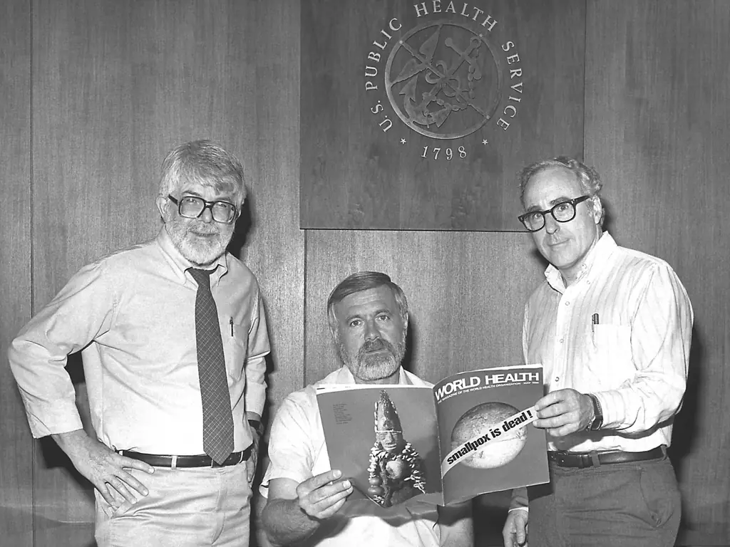
Three men in shirts and ties reading a magazine together beneath the seal of the U.S. Public Health Service.

Efforts of Eradication

By the middle of the 20th century, confidence grew that smallpox could be the first disease humankind would eradicate. In 1967 the World Health Organization launched a global smallpox eradication program. Mass vaccination of over 80 percent of each country’s population followed, but people who were nomadic or lived in politically unstable regions posed particular problems.

To reach large populations, the epidemiologist William Foege developed Eradication Escalation, which concentrated on containing outbreaks in the season when smallpox spread least, when stopping just a few cases could break a chain of transmission.

Other measures included forcibly vaccinating an Indian religious leader to convince his followers to be vaccinated, negotiating ceasefires to move vaccines through war-torn Nigeria, vaccinating in rings around each outbreak, and paying cash rewards for reporting smallpox cases. The last naturally contracted case of smallpox was reported in Somalia in 1977, and in 1980 the WHO declared smallpox eradicated.

Three men in shirts and ties reading a magazine together beneath the seal of the U.S. Public Health Service.

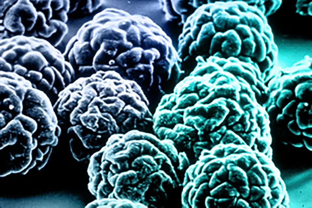
A colorized electron micrograph of virus particles.

Vaccine Innovations

Finding the germ behind a disease is only the first step. For some diseases a vaccine followed within ten years; for others the wait has lasted more than a century.

From discovering the germ to licensing a vaccine
1880190019201940196019802000MalariaMalaria: germ identified 1880; no U.S. vaccine as of 2016TuberculosisTuberculosis: germ identified 1882; no U.S. vaccine as of 2016Typhoid feverTyphoid fever: germ identified 1884; vaccine licensed 1989 (105 years)105 yrsMeningitis (Hib)Meningitis (Hib): germ identified 1889; vaccine licensed 1981 (92 years)92 yrsWhooping coughWhooping cough: germ identified 1906; vaccine licensed 1948 (42 years)42 yrsDengue feverDengue fever: germ identified 1907; no U.S. vaccine as of 2016PolioPolio: germ identified 1908; vaccine licensed 1955 (47 years)47 yrsZika feverZika fever: germ identified 1947; no U.S. vaccine as of 2016ChickenpoxChickenpox: germ identified 1953; vaccine licensed 1995 (42 years)42 yrsMeaslesMeasles: germ identified 1953; vaccine licensed 1963 (10 years)10 yrsCytomegalovirusCytomegalovirus: germ identified 1960; no U.S. vaccine as of 2016Hepatitis BHepatitis B: germ identified 1965; vaccine licensed 1981 (16 years)16 yrsRotavirus (diarrhea)Rotavirus (diarrhea): germ identified 1973; vaccine licensed 2006 (33 years)33 yrsEbolaEbola: germ identified 1976; no U.S. vaccine as of 2016Cervical cancer (HPV)Cervical cancer (HPV): germ identified 1981; vaccine licensed 2006 (25 years)25 yrsAIDS (HIV)AIDS (HIV): germ identified 1983; no U.S. vaccine as of 2016
  • Germ linked to the disease
  • Vaccine licensed in the U.S.
  • No U.S. vaccine yet (as of 2016)

Source: Our World in Data (Max Roser), CC BY-SA. Some of these diseases have gained vaccines since 2016.

A colorized electron micrograph of virus particles.

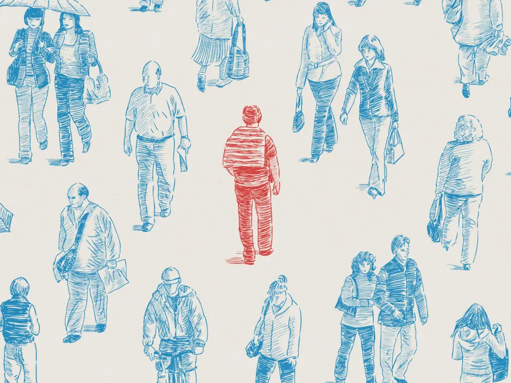
A drawing of a crowd of people in blue, with one person in red.

What Vaccines Did

Before vaccines, these diseases were part of ordinary American life. Comparing the years just before each vaccine with the years after shows how completely they changed it.

Fewer U.S. cases after each vaccine
Smallpox Smallpox: 250 cases per million a year before the vaccine (1900–49); 0 in 2006 100%Diphtheria Diphtheria: 158 cases per million a year before the vaccine (1936–45); 0 in 2006 100%Paralytic polio Paralytic polio: 103 cases per million a year before the vaccine (1951–54); 0 in 2006 100%Measles Measles: 3,044 cases per million a year before the vaccine (1953–62); 0.2 in 2006 99.99%Rubella Rubella: 242 cases per million a year before the vaccine (1966–68); 0.04 in 2006 99.98%Hib Hib: 84 cases per million a year before the vaccine (1980s); 0.17 in 2006 99.8%Mumps Mumps: 830 cases per million a year before the vaccine (1963–68); 22 in 2006 97.3%Pertussis Pertussis: 1,534 cases per million a year before the vaccine (1934–43); 52 in 2006 96.6%Tetanus Tetanus: 4 cases per million a year before the vaccine (1947–49); 0.14 in 2006 96.5%Hepatitis A Hepatitis A: 465 cases per million a year before the vaccine (1986–95); 51 in 2006 89.0%Varicella Varicella: 16,018 cases per million a year before the vaccine (1990–94); 2046 in 2006 87.2%Hepatitis B Hepatitis B: 273 cases per million a year before the vaccine (1982–91); 44 in 2006 83.9%Pneumococcal Pneumococcal: 233 cases per million a year before the vaccine (1997–99); 139 in 2006 40.3%
  • Cases a year before the vaccine
  • Cases a year in 2006
  • Reduction

Each bar is scaled to that disease’s own pre-vaccine level. Source: Roush and Murphy, Journal of the American Medical Association, 2007, via Our World in Data.

A drawing of a crowd of people in blue, with one person in red.

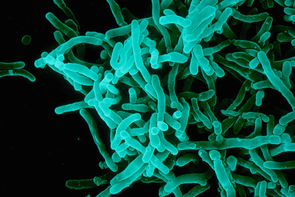
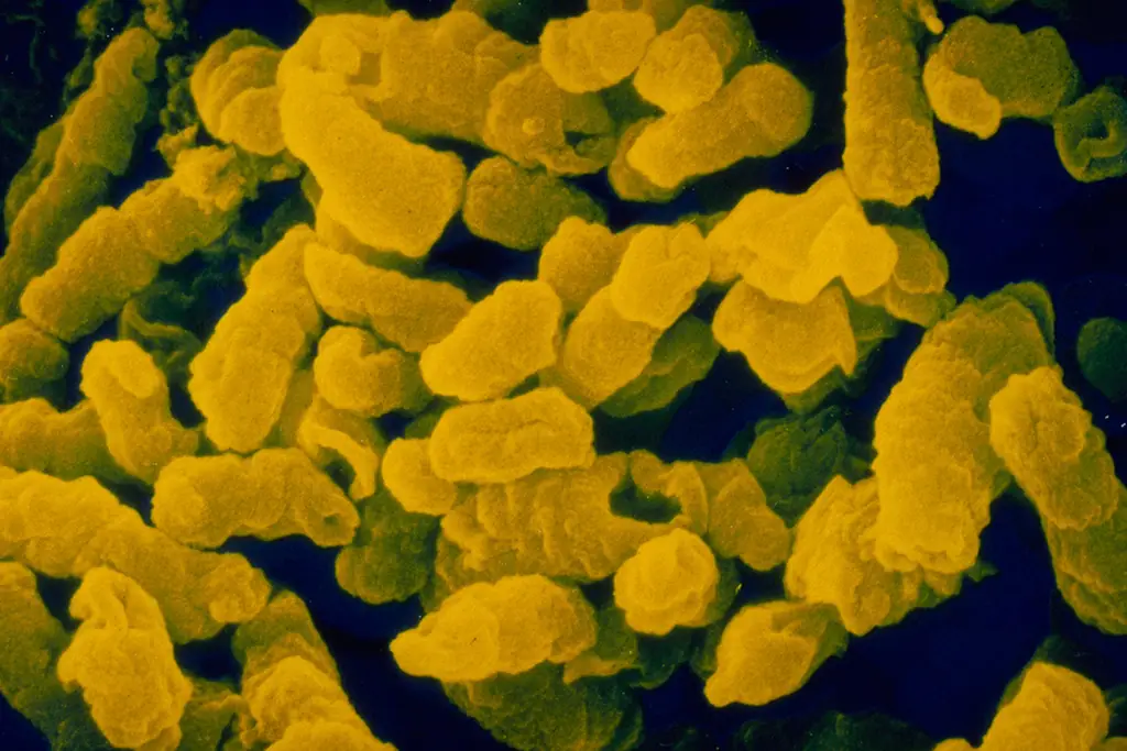
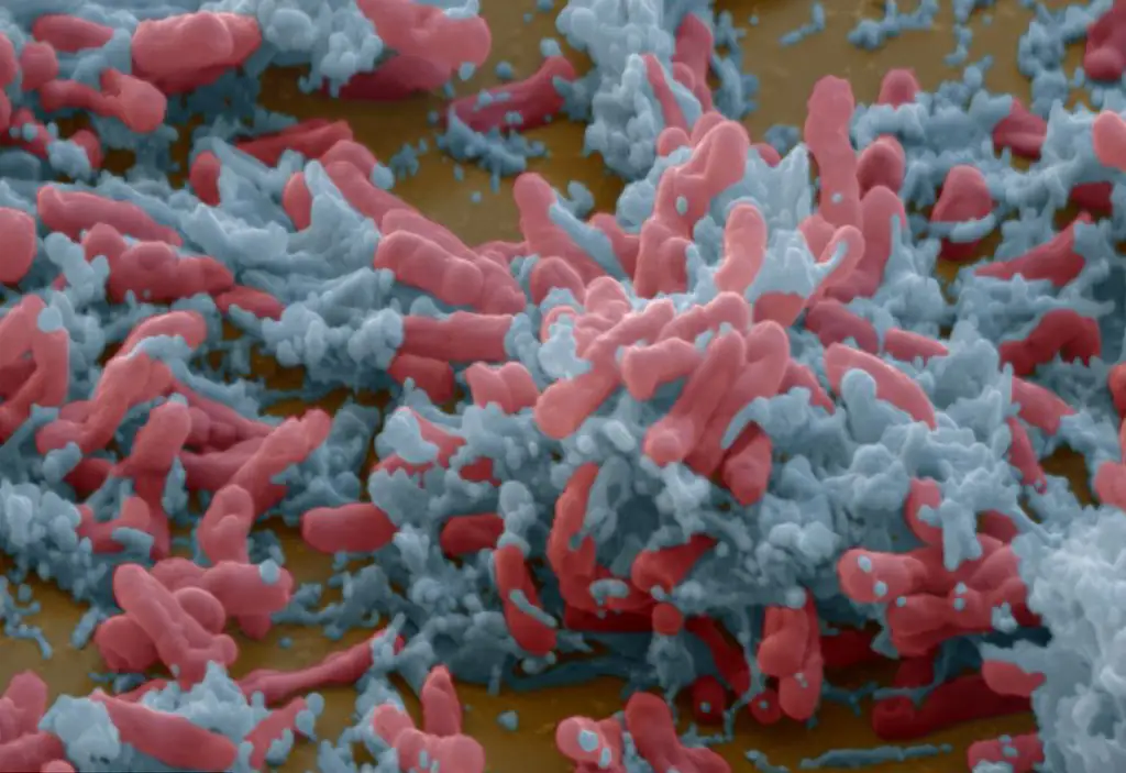
A colorized electron micrograph of rod-shaped bacteria.

Diphtheria, Tetanus and Pertussis

A colorized electron micrograph of rod-shaped bacteria.

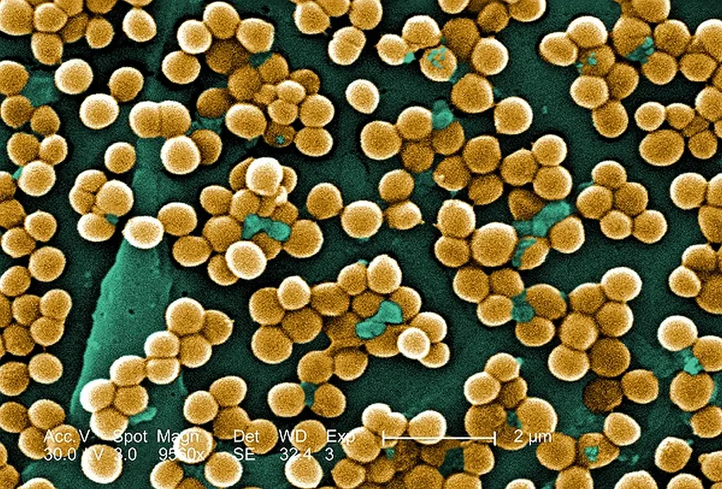
A colorized electron micrograph of clustered bacteria.

Haemophilus influenzae type b

A colorized electron micrograph of clustered bacteria.

A colorized electron micrograph of a virus particle.

Hepatitis B

A colorized electron micrograph of a virus particle.

A colorized electron micrograph of virus particles.

Poliomyelitis

A colorized electron micrograph of virus particles.

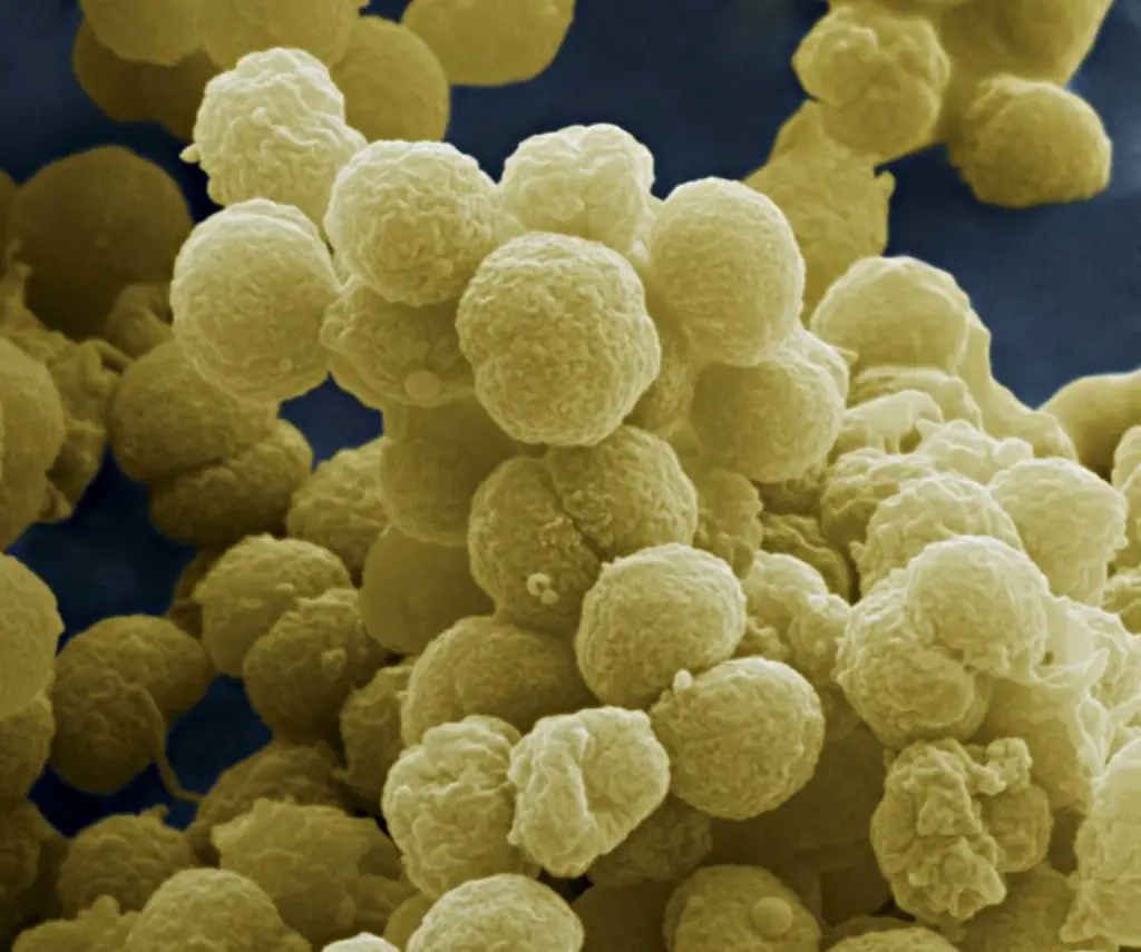
A colorized electron micrograph of round virus particles.

Measles, Mumps and Rubella

A colorized electron micrograph of round virus particles.

A colorized electron micrograph of round bacteria.

Meningococcal Meningitis

A colorized electron micrograph of round bacteria.

A colorized electron micrograph of rod-shaped bacteria.

Tuberculosis

A colorized electron micrograph of rod-shaped bacteria.

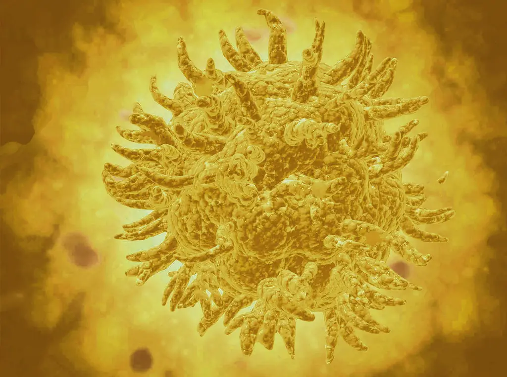
A colorized electron micrograph of a virus particle.

Yellow Fever

A colorized electron micrograph of a virus particle.

A drawing of a crowd of people in blue, with one person in red.

Herd Immunity

There is a shared benefit when more people are vaccinated. For most diseases, the greater the share of people who are immune, the better protected everyone is, because the disease can no longer spread easily. This community protection is called herd immunity.

Herd immunity especially protects people who cannot be vaccinated, such as babies who are too young, or children whose immune systems are weakened. They are the first victims when vaccination rates fall.

How many people must be immune to stop a disease
0%25%50%75%100%Share of people who must be immuneMeasles Measles (Airborne): each case infects 12–18 others; 92–95% must be immune 92–95%Pertussis Pertussis (Airborne droplet): each case infects 12–17 others; 92–94% must be immune 92–94%Diphtheria Diphtheria (Saliva): each case infects 6–7 others; 83–86% must be immune 83–86%Rubella Rubella (Airborne droplet): each case infects 6–7 others; 83–86% must be immune 83–86%Smallpox Smallpox (Airborne droplet): each case infects 5–7 others; 80–86% must be immune 80–86%Polio Polio (Fecal-oral): each case infects 5–7 others; 80–86% must be immune 80–86%Mumps Mumps (Airborne droplet): each case infects 4–7 others; 75–86% must be immune 75–86%SARS SARS (Airborne droplet): each case infects 2–5 others; 50–80% must be immune 50–80%Ebola Ebola (Bodily fluids): each case infects 1.5–2.5 others; 33–60% must be immune 33–60%Influenza Influenza (Airborne droplet): each case infects 1.5–1.8 others; 33–44% must be immune 33–44%
  • Herd immunity threshold

The more people each case infects, the higher the threshold. A measles case infects 12 to 18 people; a polio case 5 to 7.

The more contagious a disease, the higher the share of people who must be immune to stop it. Measles and whooping cough spread so easily that more than nine in ten people need to be protected. Two doses of measles vaccine give 99 percent protection; without vaccination, the lifetime risk of catching measles is nearly 100 percent.

A drawing of a crowd of people in blue, with one person in red.

Immunization

Polio Timeline

From the first clinical description of polio in 1789 to the push to wipe it out entirely. Tap an entry to read more.

  1. Loading the timeline…

Building the virus…

Explore in 3D

The poliovirus, up close

Drag to turn · pinch or scroll to zoom · tap any part

Building the model…

Explore in 3D

Drag to turn · pinch or scroll to zoom · tap any part

Polio facts